ARG45679
anti-ATP6V1A antibody
anti-ATP6V1A antibody for Flow cytometry,ICC/IF,IHC-Formalin-fixed paraffin-embedded sections,Western blot and Human,Mouse,Rat
Overview
| Product Description | Rabbit Polyclonal antibody recognizes ATP6V1A |
|---|---|
| Tested Reactivity | Hu, Ms, Rat |
| Tested Application | FACS, ICC/IF, IHC-P, WB |
| Host | Rabbit |
| Clonality | Polyclonal |
| Isotype | IgG |
| Target Name | ATP6V1A |
| Antigen Species | Human |
| Immunogen | Recombinant protein containing to human ATP6V1A. |
| Conjugation | Un-conjugated |
| Alternate Names | ATPase, H+ transporting, lysosomal 70kDa, V1 subunit A; VA68; VPP2; HO68; V-ATPase 69 kDa subunit; EC 3.6.3.14; V-ATPase subunit A; ATP6V1A1; Vma1; Vacuolar proton pump subunit alpha; ATP6A1; V-type proton ATPase catalytic subunit A; Vacuolar ATPase isoform VA68 |
Application Instructions
| Application Suggestion |
|
||||||||||
|---|---|---|---|---|---|---|---|---|---|---|---|
| Application Note | * The dilutions indicate recommended starting dilutions and the optimal dilutions or concentrations should be determined by the scientist. |
Properties
| Form | Liquid |
|---|---|
| Purification | Affinity purified |
| Buffer | 0.2% Na2HPO4, 0.9% NaCl and 4% Trehalose. |
| Stabilizer | 4% Trehalose |
| Concentration | 0.5 mg/ml |
| Storage Instruction | For continuous use, store undiluted antibody at 2-8°C for up to a week. For long-term storage, aliquot and store at -20°C or below. Storage in frost free freezers is not recommended. Avoid repeated freeze/thaw cycles. Suggest spin the vial prior to opening. The antibody solution should be gently mixed before use. |
| Note | For laboratory research only, not for drug, diagnostic or other use. |
Bioinformation
| Database Links |
Swiss-port # P38606 Human V-type proton ATPase catalytic subunit A Swiss-port # P50516 Mouse V-type proton ATPase catalytic subunit A |
|---|---|
| Gene Symbol | ATP6V1A |
| Gene Full Name | ATPase, H+ transporting, lysosomal 70kDa, V1 subunit A |
| Background | This gene encodes a component of vacuolar ATPase (V-ATPase), a multisubunit enzyme that mediates acidification of eukaryotic intracellular organelles. V-ATPase dependent organelle acidification is necessary for such intracellular processes as protein sorting, zymogen activation, receptor-mediated endocytosis, and synaptic vesicle proton gradient generation. V-ATPase is composed of a cytosolic V1 domain and a transmembrane V0 domain. The V1 domain consists of three A and three B subunits, two G subunits plus the C, D, E, F, and H subunits. The V1 domain contains the ATP catalytic site. The V0 domain consists of five different subunits: a, c, c', c", and d. Additional isoforms of many of the V1 and V0 subunit proteins are encoded by multiple genes or alternatively spliced transcript variants. This encoded protein is one of two V1 domain A subunit isoforms and is found in all tissues. Transcript variants derived from alternative polyadenylation exist. [provided by RefSeq, Jul 2008] |
| Function | Catalytic subunit of the peripheral V1 complex of vacuolar ATPase. V-ATPase vacuolar ATPase is responsible for acidifying a variety of intracellular compartments in eukaryotic cells. [UniProt] |
| Cellular Localization | Cytoplasm; Cytoplasmic vesicle; Lysosome; Membrane. [UniProt] |
| Calculated MW | 68 kDa |
| PTM | Acetylation; Phosphoprotein. [UniProt] |
Images (4) Click the Picture to Zoom In
-
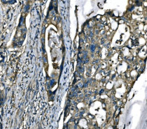
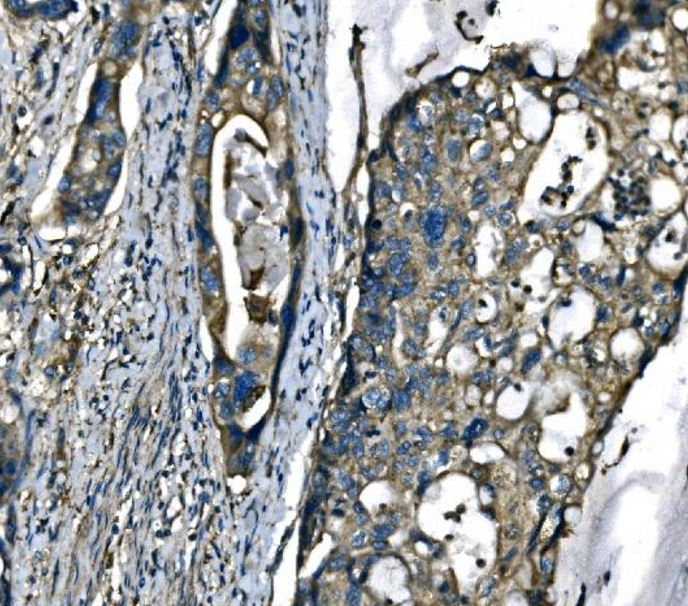
ARG45679 anti-ATP6V1A antibody IHC-P image
Immunohistochemistry: Human adenocarcinoma of the right colon stained with ARG45679 anti-ATP6V1A antibody at 2 μg/ml dilution.
-
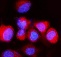
ARG45679 anti-ATP6V1A antibody ICC/IF image
Immunofluorescence: A549 stained with ARG45679 anti-ATP6V1A antibody at 5 μg/ml dilution.
-
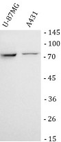
ARG45679 anti-ATP6V1A antibody WB image
Western blot: U-87MG and A431 stained with ARG45679 anti-ATP6V1A antibody at 0.5 μg/ml dilution.
-
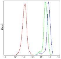
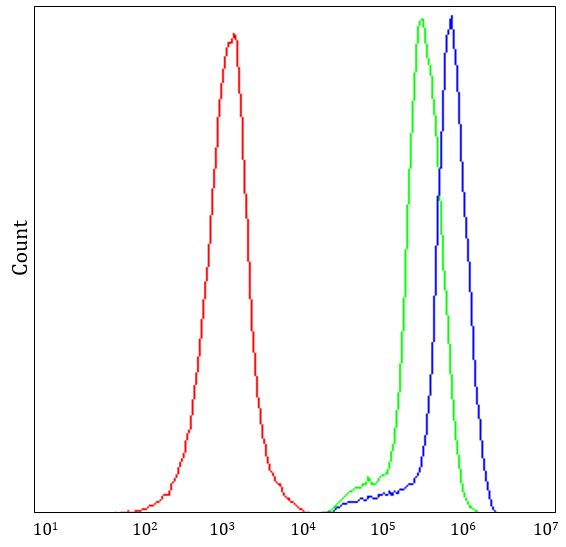
ARG45679 anti-ATP6V1A antibody FACS image
Flow Cytometry: JK stained with ARG45679 anti-ATP6V1A antibody at 1 µg/10^6 cells dilution.