ARG46039
anti-BCL2L10 antibody
anti-BCL2L10 antibody for Western blot and Human
Overview
| Product Description | Rabbit Polyclonal antibody recognizes BCL2L10 |
|---|---|
| Tested Reactivity | Hu |
| Tested Application | WB |
| Host | Rabbit |
| Clonality | Polyclonal |
| Isotype | IgG |
| Target Name | BCL2L10 |
| Antigen Species | Human |
| Immunogen | A 16 amino acid synthetic peptide within aa. 90 - 140 of human BCL2L10. |
| Conjugation | Un-conjugated |
| Alternate Names | BCL2L10; BCL2-like 10 (apoptosis facilitator); Bcl-B; Boo; Diva; BCL-B; BCLB; Bcl-2-like protein 10; Anti-apoptotic protein NrH; Bcl2-L-10 |
Application Instructions
| Application Suggestion |
|
||||
|---|---|---|---|---|---|
| Application Note | * The dilutions indicate recommended starting dilutions and the optimal dilutions or concentrations should be determined by the scientist. | ||||
| Observed Size | 26 kDa |
Properties
| Purification | Affinity chromatography purified |
|---|---|
| Buffer | PBS and 0.02% Sodium azide. |
| Preservative | 0.02% Sodium azide |
| Concentration | 1 mg/ml |
| Storage Instruction | For continuous use, store undiluted antibody at 2-8°C for up to a week. For long-term storage, aliquot and store at -89°C or below. Storage in frost free freezers is not recommended. Avoid repeated freeze/thaw cycles. Suggest spin the vial prior to opening. The antibody solution should be gently mixed before use. |
| Note | For laboratory research only, not for drug, diagnostic or other use. |
Bioinformation
| Database Links | |
|---|---|
| Gene Symbol | BCL2L10 |
| Gene Full Name | BCL2-like 10 (apoptosis facilitator) |
| Background | The protein encoded by this gene belongs to the BCL-2 protein family. BCL-2 family members form hetero- or homodimers and act as anti- or pro-apoptotic regulators that are involved in a wide variety of cellular activities. The protein encoded by this gene contains conserved BH4, BH1 and BH2 domains. This protein can interact with other members of BCL-2 protein family including BCL2, BCL2L1/BCL-X(L), and BAX. Overexpression of this gene has been shown to suppress cell apoptosis possibly through the prevention of cytochrome C release from the mitochondria, and thus activating caspase-3 activation. The mouse counterpart of this protein is found to interact with Apaf1 and forms a protein complex with Caspase 9, which suggests the involvement of this protein in APAF1 and CASPASE 9 related apoptotic pathway. [provided by RefSeq, Jul 2008] |
| Function | Promotes cell survival by suppressing apoptosis induced by BAX but not BAK (PubMed:11278245, PubMed:11689480). Increases binding of AHCYL1/IRBIT to ITPR1 (PubMed:27995898). Reduces ITPR1-mediated calcium release from the endoplasmic reticulum cooperatively with AHCYL1/IRBIT under normal cellular conditions (PubMed:27995898). Under apoptotic stress conditions, dissociates from ITPR1 and is displaced from mitochondria-associated endoplasmic reticulum membranes, leading to increased Ca(2+) transfer to mitochondria which promotes apoptosis (PubMed:27995898). Required for the correct formation of the microtubule organizing center during oocyte cell division, potentially via regulation of protein abundance and localization of other microtubule organizing center components such as AURKA and TPX2 (By similarity). [UniProt] |
| Cellular Localization | Mitochondrion. [UniProt] |
| Calculated MW | 22 kDa |
| PTM | Phosphoprotein. [UniProt] |
Images (1) Click the Picture to Zoom In
-
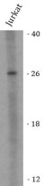
ARG46039 anti-BCL2L10 antibody WB image
Western blot: Jurkat stained with ARG46039 anti-BCL2L10 antibody.


