ARG55068
anti-Caveolin 2 antibody
anti-Caveolin 2 antibody for Flow cytometry,ICC/IF,IHC-Formalin-fixed paraffin-embedded sections,Western blot and Human
Cell Biology and Cellular Response antibody; Cell Death antibody; Controls and Markers antibody

Overview
| Product Description | Rabbit Polyclonal antibody recognizes Caveolin 2 |
|---|---|
| Tested Reactivity | Hu |
| Tested Application | FACS, ICC/IF, IHC-P, WB |
| Host | Rabbit |
| Clonality | Polyclonal |
| Isotype | IgG |
| Target Name | Caveolin 2 |
| Antigen Species | Human |
| Immunogen | KLH-conjugated synthetic peptide corresponding to aa. 11-44 (N-terminus) of Human Caveolin 2. |
| Conjugation | Un-conjugated |
| Alternate Names | Caveolin-2; CAV |
Application Instructions
| Application Suggestion |
|
||||||||||
|---|---|---|---|---|---|---|---|---|---|---|---|
| Application Note | * The dilutions indicate recommended starting dilutions and the optimal dilutions or concentrations should be determined by the scientist. |
Properties
| Form | Liquid |
|---|---|
| Purification | Saturated Ammonium Sulfate (SAS) precipitation followed by dialysis against PBS. |
| Buffer | PBS and 0.09% (W/V) Sodium azide |
| Preservative | 0.09% (W/V) Sodium azide |
| Storage Instruction | For continuous use, store undiluted antibody at 2-8°C for up to a week. For long-term storage, aliquot and store at -20°C or below. Storage in frost free freezers is not recommended. Avoid repeated freeze/thaw cycles. Suggest spin the vial prior to opening. The antibody solution should be gently mixed before use. |
| Note | For laboratory research only, not for drug, diagnostic or other use. |
Bioinformation
| Database Links | |
|---|---|
| Gene Symbol | CAV2 |
| Gene Full Name | caveolin 2 |
| Background | The protein encoded by this gene is a major component of the inner surface of caveolae, small invaginations of the plasma membrane, and is involved in essential cellular functions, including signal transduction, lipid metabolism, cellular growth control and apoptosis. This protein may function as a tumor suppressor. This gene and related family member (CAV1) are located next to each other on chromosome 7, and express colocalizing proteins that form a stable hetero-oligomeric complex. Alternatively spliced transcript variants encoding different isoforms have been identified for this gene. Additional isoforms resulting from the use of alternate in-frame translation initiation codons have also been described, and shown to have preferential localization in the cell (PMID:11238462). [provided by RefSeq, May 2011] |
| Function | May act as a scaffolding protein within caveolar membranes. Interacts directly with G-protein alpha subunits and can functionally regulate their activity. Acts as an accessory protein in conjunction with CAV1 in targeting to lipid rafts and driving caveolae formation. The Ser-36 phosphorylated form has a role in modulating mitosis in endothelial cells. Positive regulator of cellular mitogenesis of the MAPK signaling pathway. Required for the insulin-stimulated nuclear translocation and activation of MAPK1 and STAT3, and the subsequent regulation of cell cycle progression (By similarity). [UniProt] |
| Cellular Localization | Cell membrane, Cytoplasm, Golgi apparatus, Membrane, Nucleus |
| Research Area | Cell Biology and Cellular Response antibody; Cell Death antibody; Controls and Markers antibody |
| Calculated MW | 18 kDa |
| PTM | Phosphorylated on serine and tyrosine residues. CAV1 promotes phosphorylation on Ser-23 which then targets the complex to the plasma membrane, lipid rafts and caveolae. Phosphorylation on Ser-36 appears to modulate mitosis in endothelial cells (By similarity). Phosphorylation on both Tyr-19 and Tyr-27 is required for insulin-induced 'Ser-727' phosphorylation of STAT3 and its activation. Phosphorylation on Tyr-19 is required for insulin-induced phosphorylation of MAPK1 and DNA binding of STAT3. Tyrosine phosphorylation is induced by both EGF and insulin (By similarity). |
Images (4) Click the Picture to Zoom In
-
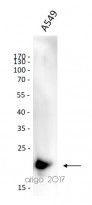
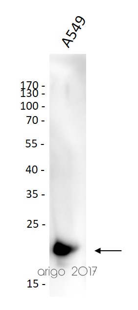
ARG55068 anti-Caveolin 2 antibody WB image
Western blot: 30 µg of A549 cell lysate stained with ARG55068 anti-Caveolin 2 antibody at 1:1000 dilution.
-
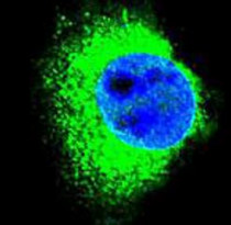
ARG55068 anti-Caveolin 2 antibody ICC/IF image
Immunofluorescence: MDA-MB-231 cells stained with ARG55068 anti-Caveolin 2 antibody (green). DAPI (blue) for nuclear staining.
-
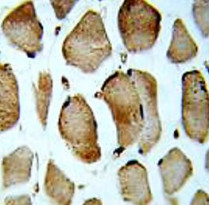
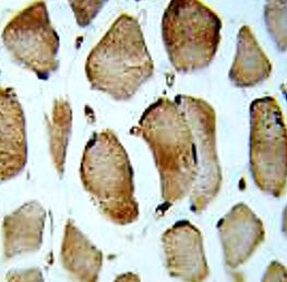
ARG55068 anti-Caveolin 2 antibody IHC-P image
Immunohistochemistry: Formalin-fixed and paraffin-embedded Human skeletal muscle stained with ARG55068 anti-Caveolin 2 antibody.
-

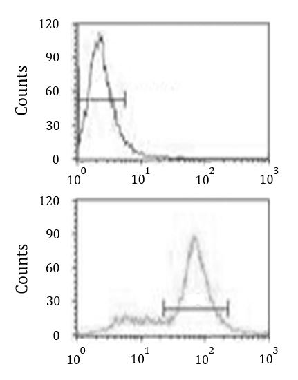
ARG55068 anti-Caveolin 2 antibody FACS image
Flow Cytometry: MDA-MB231 cells stained with ARG55068 anti-Caveolin 2 antibody (bottom histogram) or without primary antibody control (top histogram), followed by incubation with FITC labelled secondary antibody.

Customer's Feedback
Excellent
Review for anti-Caveolin 2 antibody
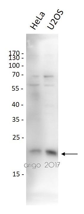
Application:WB
Sample:HeLa and U2OS
Sample Loading Amount:30 µg
Primary Antibody Dilution Factor:1:1000
Primary Antibody Incubation Time:overnight
Primary Antibody Incubation Temperature:4 ºC


