ARG45564
anti-Cofilin antibody
anti-Cofilin antibody for Flow cytometry,ICC/IF,IHC-Formalin-fixed paraffin-embedded sections,IHC-Frozen sections,Western blot and Human,Monkey,Mouse,Rat
Overview
| Product Description | Rabbit Polyclonal antibody recognizes Cofilin |
|---|---|
| Tested Reactivity | Hu, Ms, Rat, Mk |
| Tested Application | FACS, ICC/IF, IHC-Fr, IHC-P, WB |
| Host | Rabbit |
| Clonality | Polyclonal |
| Isotype | IgG |
| Target Name | Cofilin |
| Antigen Species | Human |
| Immunogen | Recombinant protein containing to human Cofilin. |
| Conjugation | Un-conjugated |
| Alternate Names | CFL1; cofilin 1 (non-muscle); CFL; 18 kDa phosphoprotein; Cofilin, non-muscle isoform; HEL-S-15; p18; cofilin; Cofilin-1 |
Application Instructions
| Application Suggestion |
|
||||||||||||
|---|---|---|---|---|---|---|---|---|---|---|---|---|---|
| Application Note | * The dilutions indicate recommended starting dilutions and the optimal dilutions or concentrations should be determined by the scientist. | ||||||||||||
| Observed Size | 19 kDa |
Properties
| Form | Liquid |
|---|---|
| Purification | Affinity purified |
| Buffer | 0.9% NaCl, 0.2% Na2HPO4, 0.05% Sodium azide and 5% BSA. |
| Preservative | 0.05% Sodium azide |
| Stabilizer | 5% BSA |
| Concentration | 0.5 mg/ml |
| Storage Instruction | For continuous use, store undiluted antibody at 2-8°C for up to a week. For long-term storage, aliquot and store at -20°C or below. Storage in frost free freezers is not recommended. Avoid repeated freeze/thaw cycles. Suggest spin the vial prior to opening. The antibody solution should be gently mixed before use. |
| Note | For laboratory research only, not for drug, diagnostic or other use. |
Bioinformation
| Database Links | |
|---|---|
| Gene Symbol | CFL1 |
| Gene Full Name | cofilin 1 (non-muscle) |
| Background | Members of the ADF/cofilin (AC) family are actin-severing proteins that regulate actin remodeling during cellular events such as cell migration, cytokinesis, phagocytosis, endocytosis, axon development, and immune cell activation. In mammals, there are three members of the AC family, muscle-specific cofilin (Cofilin 2), non-muscle cofilin (Cofilin 1), and ADF. In humans, cofilin 1 and ADF have 72% identity, with the major amino acid differences found in the C-terminal region. Regulation of cofilin activity can occur through serine phosphorylation. Activation of cofilin kinases, LIMK1 or LIMK2, leads to phosphorylation of cofilin at serine 3. This phosphorylation disrupts cofilin binding to actin in vitro and in vivo. Multiple phosphatases, PP1, PP2A, PP2B, slingshot, and chronophin can dephosphorylate Ser-3 and activate actin binding. Thus, Ser-3 phosphorylation is a major site for the regulation of cofilin activity. |
| Function | Binds to F-actin and exhibits pH-sensitive F-actin depolymerizing activity. Regulates actin cytoskeleton dynamics. Important for normal progress through mitosis and normal cytokinesis. Plays a role in the regulation of cell morphology and cytoskeletal organization. Required for the up-regulation of atypical chemokine receptor ACKR2 from endosomal compartment to cell membrane, increasing its efficiency in chemokine uptake and degradation. [UniProt] |
| Cellular Localization | Cell membrane; Cell projection; Cytoplasm; Cytoskeleton; Membrane; Nucleus. [UniProt] |
| Calculated MW | 19 kDa |
| PTM | Inactivated by phosphorylation on Ser-3. Phosphorylated on Ser-3 in resting cells (By similarity). Dephosphorylated by PDXP/chronophin; this restores its activity in promoting actin filament depolymerization. The phosphorylation of Ser-24 may prevent recognition of the nuclear localization signal (By similarity). Phosphorylated via a ARRB1-RAC1-LIMK1-PAK1 cascade upon active ligand stimulation of atypical chemokine receptor ACKR2.. [UniProt] |
Images (10) Click the Picture to Zoom In
-
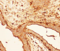
ARG45564 anti-Cofilin antibody IHC-P image
Immunohistochemistry: Human intestinal cancer stained with ARG45564 anti-Cofilin antibody at 1 μg/ml dilution.
-
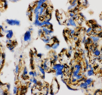
ARG45564 anti-Cofilin antibody IHC-Fr image
Immunohistochemistry: Frozen Human placenta stained with ARG45564 anti-Cofilin antibody at 1 μg/ml dilution.
-
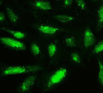
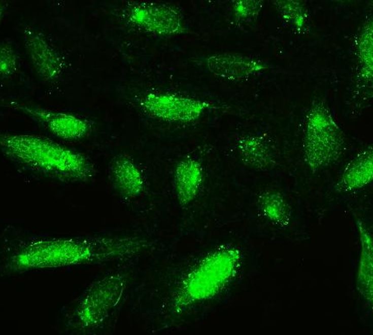
ARG45564 anti-Cofilin antibody ICC/IF image
Immunofluorescence: U2OS stained with ARG45564 anti-Cofilin antibody at 5 μg/ml dilution.
-
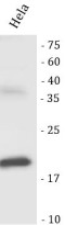
ARG45564 anti-Cofilin antibody WB image
Western blot: Hela stained with ARG45564 anti-Cofilin antibody at 0.5 μg/ml dilution.
-
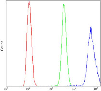
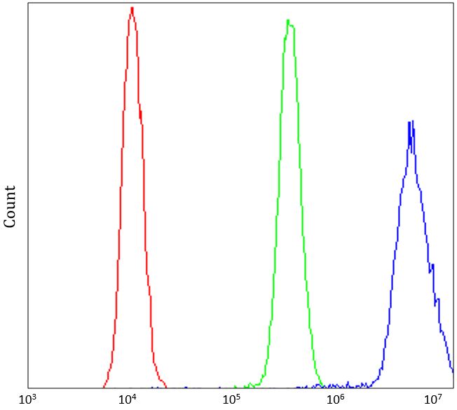
ARG45564 anti-Cofilin antibody FACS image
Flow Cytometry: U2OS stained with ARG45564 anti-Cofilin antibody at 1 µg/10^6 cells dilution.
-
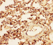
ARG45564 anti-Cofilin antibody IHC-P image
Immunohistochemistry: Rat lung stained with ARG45564 anti-Cofilin antibody at 1 μg/ml dilution.
-
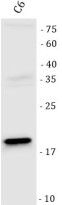
ARG45564 anti-Cofilin antibody WB image
Western blot: C6 stained with ARG45564 anti-Cofilin antibody at 0.5 μg/ml dilution.
-
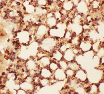
ARG45564 anti-Cofilin antibody IHC-P image
Immunohistochemistry: Mouse lung stained with ARG45564 anti-Cofilin antibody at 1 μg/ml dilution.
-
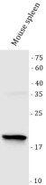
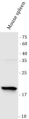
ARG45564 anti-Cofilin antibody WB image
Western blot: Mouse spleen stained with ARG45564 anti-Cofilin antibody at 0.5 μg/ml dilution.
-

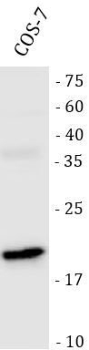
ARG45564 anti-Cofilin antibody WB image
Western blot: COS-7 stained with ARG45564 anti-Cofilin antibody at 0.5 μg/ml dilution.