ARG10354
anti-D-Dimer antibody [DD1]
anti-D-Dimer antibody [DD1] for ELISA,Immunoassay,Western blot and Human
Cell Biology and Cellular Response antibody
Overview
| Product Description | Mouse Monoclonal antibody [DD1] recognizes D-Dimer |
|---|---|
| Tested Reactivity | Hu |
| Tested Application | ELISA, IA, WB |
| Specificity | Do not cross-react with fibrinogen. |
| Host | Mouse |
| Clonality | Monoclonal |
| Clone | DD1 |
| Isotype | IgG2a |
| Target Name | D-Dimer |
| Antigen Species | Mouse |
| Immunogen | homogenized fibrin clot, D-dimer or high molecular weight fibrin degradation products. |
| Conjugation | Un-conjugated |
Application Instructions
| Application Note | * The dilutions indicate recommended starting dilutions and the optimal dilutions or concentrations should be determined by the scientist. |
|---|
Properties
| Form | Liquid |
|---|---|
| Purification | Protein A affinity purified. |
| Buffer | PBS (pH 7.4) and 0.1% Sodium azide |
| Preservative | 0.1% Sodium azide |
| Concentration | 1.0-2.0 mg/ml |
| Storage Instruction | For continuous use, store undiluted antibody at 2-8°C for up to a week. For long-term storage, aliquot and store at -20°C or below. Storage in frost free freezers is not recommended. Avoid repeated freeze/thaw cycles. Suggest spin the vial prior to opening. The antibody solution should be gently mixed before use. |
| Note | For laboratory research only, not for drug, diagnostic or other use. |
Bioinformation
| Research Area | Cell Biology and Cellular Response antibody |
|---|
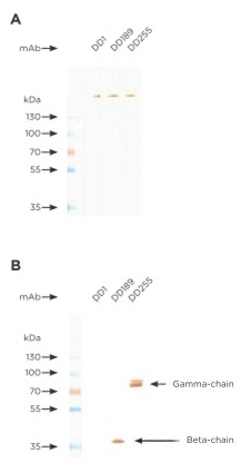
Images (1) Click the Picture to Zoom In
-
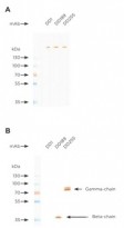
ARG10354 anti-D-Dimer antibody [DD1] WB image
Western Blot: D-dimer was run in SDS-PAGE under non-reducing (A) or reducing (B) conditions. Using a 7.5–12.5% separating gel and transferred onto a nitrocellulose membrane.
The membrane was blocked by 7% milk in PBST for 30 minutes and the protein bands were stained by different 4 D-dimer mAbs (10 µg/ml) 1) anti-D-Dimer antibody [DD1] (ARG10354); 2) anti-D-Dimer antibody [DD189]; 3) anti-D-Dimer antibody [DD255] stained with anti-D-Dimer antibody [DD1] (ARG10354) for 1 hour. After washing with PBST, goat anti-mouse Fc-specific IgG labeled with horseradish peroxidase was added and incubated for 1 hour. After washing with PBST, the immune complexes were visualized by DAB/hydrogen peroxide in 50 mM Tris-HCl buffer, pH7.5.
Clone References
Protein biomarkers in exfoliated cells collected from the human rectal mucosa: implications for colorectal disease detection and monitoring.
ELISA / Human


Activation of mannan-binding lectin-associated serine proteases leads to generation of a fibrin clot.
Activation of plasminogen activator inhibitor implicates protease InhA in the acute-phase response to Bacillus anthracis infection.
ELISA /
Neutrophil elastase and syndecan shedding contribute to antithrombin depletion in murine anthrax.


