ARG63442
anti-DBNL / HIP55 antibody
anti-DBNL / HIP55 antibody for Western blot and Human
Signaling Transduction antibody
Overview
| Product Description | Goat Polyclonal antibody recognizes DBNL / HIP55 |
|---|---|
| Tested Reactivity | Hu |
| Predict Reactivity | Ms, Rat, Dog |
| Tested Application | WB |
| Specificity | This antibody is expected to recognise all reported isoforms (NP_054782.2; NP_001014436.1; NP_001116428.1) |
| Host | Goat |
| Clonality | Polyclonal |
| Isotype | IgG |
| Target Name | DBNL / HIP55 |
| Antigen Species | Human |
| Immunogen | AANLSRNGPALQE-C |
| Conjugation | Un-conjugated |
| Alternate Names | SH3P7; HIP55; SH3 domain-containing protein 7; ABP1; Cervical SH3P7; Drebrin-F; HPK1-interacting protein of 55 kDa; Drebrin-like protein; HIP-55; Cervical mucin-associated protein |
Application Instructions
| Application Suggestion |
|
||||
|---|---|---|---|---|---|
| Application Note | WB: Recommend incubate at RT for 1h. * The dilutions indicate recommended starting dilutions and the optimal dilutions or concentrations should be determined by the scientist. |
Properties
| Form | Liquid |
|---|---|
| Purification | Purified from goat serum by antigen affinity chromatography. |
| Buffer | Tris saline (pH 7.3), 0.02% Sodium azide and 0.5% BSA. |
| Preservative | 0.02% Sodium azide |
| Stabilizer | 0.5% BSA |
| Concentration | 0.5 mg/ml |
| Storage Instruction | For continuous use, store undiluted antibody at 2-8°C for up to a week. For long-term storage, aliquot and store at -20°C or below. Storage in frost free freezers is not recommended. Avoid repeated freeze/thaw cycles. Suggest spin the vial prior to opening. The antibody solution should be gently mixed before use. |
| Note | For laboratory research only, not for drug, diagnostic or other use. |
Bioinformation
| Database Links | |
|---|---|
| Gene Symbol | DBNL |
| Gene Full Name | drebrin-like |
| Function | Adapter protein that binds F-actin and DNM1, and thereby plays a role in receptor-mediated endocytosis. Plays a role in the reorganization of the actin cytoskeleton, formation of cell projections, such as neurites, in neuron morphogenesis and synapse formation via its interaction with WASL and COBL. Does not bind G-actin and promote actin polymerization by itself. Required for the formation of organized podosome rosettes (By similarity). May act as a common effector of antigen receptor-signaling pathways in leukocytes. Acts as a key component of the immunological synapse that regulates T-cell activation by bridging TCRs and the actin cytoskeleton to gene activation and endocytic processes. [UniProt] |
| Research Area | Signaling Transduction antibody |
| Calculated MW | 48 kDa |
| PTM | Degraded by caspases during apoptosis. |
Images (2) Click the Picture to Zoom In
-

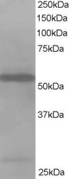
ARG63442 anti-DBNL / HIP55 antibody WB image
Western blot: Jurkat lysate (RIPA buffer, 35 µg total protein per lane) stained with ARG63442 anti-DBNL / HIP55 antibody at 1 µg/ml dilution.
-

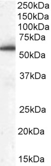
ARG63442 anti-DBNL / HIP55 antibody WB image
Western blot: 35 µg of Human peripheral blood mononucleocyte lysate (in RIPA buffer) stained with ARG63442 anti-DBNL / HIP55 antibody at 0.03 µg/ml dilution and incubated at RT for 1 hour.