ARG59557
anti-E2F1 antibody
anti-E2F1 antibody for Flow cytometry,ICC/IF,IHC-Formalin-fixed paraffin-embedded sections,Immunoprecipitation,Western blot and Human,Mouse,Rat
Overview
| Product Description | Rabbit Polyclonal antibody recognizes E2F1 |
|---|---|
| Tested Reactivity | Hu, Ms, Rat |
| Tested Application | FACS, ICC/IF, IHC-P, IP, WB |
| Host | Rabbit |
| Clonality | Polyclonal |
| Isotype | IgG |
| Target Name | E2F1 |
| Antigen Species | Human |
| Immunogen | Synthetic peptide derived from Human E2F1. |
| Conjugation | Un-conjugated |
| Alternate Names | RBAP1; Retinoblastoma-associated protein 1; Retinoblastoma-binding protein 3; RBBP3; pRB-binding protein E2F-1; RBBP-3; E2F-1; Transcription factor E2F1; RBP3; PBR3; RBAP-1 |
Application Instructions
| Application Suggestion |
|
||||||||||||
|---|---|---|---|---|---|---|---|---|---|---|---|---|---|
| Application Note | * The dilutions indicate recommended starting dilutions and the optimal dilutions or concentrations should be determined by the scientist. | ||||||||||||
| Positive Control | HeLa |
Properties
| Form | Liquid |
|---|---|
| Purification | Affinity purified. |
| Buffer | PBS (pH 7.4), 0.02% Sodium azide and 50% Glycerol. |
| Preservative | 0.02% Sodium azide |
| Stabilizer | 50% Glycerol |
| Storage Instruction | For continuous use, store undiluted antibody at 2-8°C for up to a week. For long-term storage, aliquot and store at -20°C. Storage in frost free freezers is not recommended. Avoid repeated freeze/thaw cycles. Suggest spin the vial prior to opening. The antibody solution should be gently mixed before use. |
| Note | For laboratory research only, not for drug, diagnostic or other use. |
Bioinformation
| Database Links | |
|---|---|
| Gene Symbol | E2F1 |
| Gene Full Name | E2F transcription factor 1 |
| Background | The protein encoded by this gene is a member of the E2F family of transcription factors. The E2F family plays a crucial role in the control of cell cycle and action of tumor suppressor proteins and is also a target of the transforming proteins of small DNA tumor viruses. The E2F proteins contain several evolutionally conserved domains found in most members of the family. These domains include a DNA binding domain, a dimerization domain which determines interaction with the differentiation regulated transcription factor proteins (DP), a transactivation domain enriched in acidic amino acids, and a tumor suppressor protein association domain which is embedded within the transactivation domain. This protein and another 2 members, E2F2 and E2F3, have an additional cyclin binding domain. This protein binds preferentially to retinoblastoma protein pRB in a cell-cycle dependent manner. It can mediate both cell proliferation and p53-dependent/independent apoptosis. [provided by RefSeq, Jul 2008] |
| Function | Transcription activator that binds DNA cooperatively with DP proteins through the E2 recognition site, 5'-TTTC[CG]CGC-3' found in the promoter region of a number of genes whose products are involved in cell cycle regulation or in DNA replication. The DRTF1/E2F complex functions in the control of cell-cycle progression from G1 to S phase. E2F1 binds preferentially RB1 in a cell-cycle dependent manner. It can mediate both cell proliferation and TP53/p53-dependent apoptosis. Blocks adipocyte differentiation by binding to specific promoters repressing CEBPA binding to its target gene promoters. [UniProt] |
| Cellular Localization | Nucleus. [UniProt] |
| Calculated MW | 47 kDa |
| PTM | Phosphorylated by CDK2 and cyclin A-CDK2 in the S-phase. Phosphorylation at Ser-364 by CHEK2 stabilizes E2F1 upon DNA damage and regulates its effect on transcription and apoptosis. Acetylation stimulates DNA-binding. Enhanced under stress conditions such as DNA damage and inhibited by retinoblastoma protein RB1. Regulated by KAP1/TRIM28 which recruits HDAC1 to E2F1 resulting in deacetylation. Acetylated by P/CAF/KAT2B. [UniProt] |
Images (4) Click the Picture to Zoom In
-

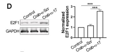
ARG59557 anti-E2F1 antibody WB image
Western blot: MCF-7 stained with ARG59557 anti-E2F1 antibody.
From Chiglintseva D et al. Biomaterials. (2024), doi: 10.1016/j.biomaterials.2024.122604, Fig. 6. D.
-
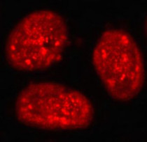
ARG59557 anti-E2F1 antibody ICC/IF image
Immunofluorescence: HeLa cells stained with ARG59557 anti-E2F1 antibody.
-

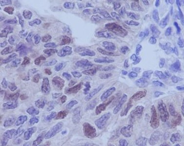
ARG59557 anti-E2F1 antibody IHC-P image
Immunohistochemistry: Paraffin-embedded Human ovary stained with ARG59557 anti-E2F1 antibody.
-
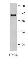
ARG59557 anti-E2F1 antibody WB image
Western blot: HeLa cell lysate stained with ARG59557 anti-E2F1 antibody.


Specific References
Targeted Inhibition of Oncogenic microRNAs miR-21, miR-17, and miR-155 Suppresses Tumor Growth and Modulates Immune Response in Colorectal Cancer
WB / Mouse


Engineering supramolecular dynamics of self-assembly and turnover of oncogenic microRNAs to drive their synergistic destruction in tumor models
WB / Human


