ARG46726
anti-LC3A/B antibody
anti-LC3A/B antibody for ICC/IF,IHC-Formalin-fixed paraffin-embedded sections,Western blot and Human,Mouse,Rat
Overview
| Product Description | Rabbit Polyclonal antibody recognizes LC3A/B |
|---|---|
| Tested Reactivity | Hu, Ms, Rat |
| Tested Application | ICC/IF, IHC-P, WB |
| Host | Rabbit |
| Clonality | Polyclonal |
| Isotype | IgG |
| Target Name | LC3A/B |
| Conjugation | Un-conjugated |
| Alternate Names | MAP1A/MAP1B light chain 3 A; MAP1BLC3; LC3; MAP1 light chain 3-like protein 1; MAP1A/MAP1B LC3 A; Autophagy-related protein LC3 A; Microtubule-associated protein 1 light chain 3 alpha; MAP1ALC3; Microtubule-associated proteins 1A/1B light chain 3A; LC3A; ATG8E; Autophagy-related ubiquitin-like modifier LC3 A; Microtubule-associated proteins 1A/1B light chain 3B; MAP1A/MAP1B light chain 3 B; MAP1A/1BLC3; MAP1 light chain 3-like protein 2; Autophagy-related protein LC3 B; MAP1A/MAP1B LC3 B; LC3B; MAP1LC3B-a; ATG8F; Microtubule-associated protein 1 light chain 3 beta; Autophagy-related ubiquitin-like modifier LC3 B |
Application Instructions
| Application Suggestion |
|
||||||||
|---|---|---|---|---|---|---|---|---|---|
| Application Note | * The dilutions indicate recommended starting dilutions and the optimal dilutions or concentrations should be determined by the scientist. |
Properties
| Form | Liquid |
|---|---|
| Purification | Affinity chromatography purified |
| Buffer | PBS, 0.09% Sodium azide and 50% Glycerol. |
| Preservative | 0.09% Sodium azide |
| Stabilizer | 50% Glycerol |
| Storage Instruction | For continuous use, store undiluted antibody at 2-8°C for up to a week. For long-term storage, aliquot and store at -20°C or below. Storage in frost free freezers is not recommended. Avoid repeated freeze/thaw cycles. Suggest spin the vial prior to opening. The antibody solution should be gently mixed before use. |
| Note | For laboratory research only, not for drug, diagnostic or other use. |
Bioinformation
| Gene Symbol | LC3A/LC3B |
|---|---|
| Gene Full Name | microtubule-associated protein 1 light chain 3 alpha / microtubule-associated protein 1 light chain 3 beta |
| Background | MAP1A and MAP1B are microtubule-associated proteins which mediate the physical interactions between microtubules and components of the cytoskeleton. MAP1A and MAP1B each consist of a heavy chain subunit and multiple light chain subunits. The protein encoded by this gene is one of the light chain subunits and can associate with either MAP1A or MAP1B. Two transcript variants encoding different isoforms have been found for this gene. The expression of variant 1 is suppressed in many tumor cell lines, suggesting that may be involved in carcinogenesis. [provided by RefSeq, Feb 2012] LC3B is a subunit of neuronal microtubule-associated MAP1A and MAP1B proteins, which are involved in microtubule assembly and important for neurogenesis. Studies on the rat homolog implicate a role for this gene in autophagy, a process that involves the bulk degradation of cytoplasmic component. [provided by RefSeq, Jul 2008] |
| Function | Ubiquitin-like modifier involved in formation of autophagosomal vacuoles (autophagosomes). Whereas LC3s are involved in elongation of the phagophore membrane, the GABARAP/GATE-16 subfamily is essential for a later stage in autophagosome maturation. [From Uniprot] |
| Calculated MW | 14 kDa, 16 kDa |
| PTM | The precursor molecule is cleaved by ATG4B to form the cytosolic form, LC3-I. This is activated by APG7L/ATG7, transferred to ATG3 and conjugated to phospholipid to form the membrane-bound form, LC3-II (PubMed:15187094). The Legionella effector RavZ is a deconjugating enzyme that produces an ATG8 product that would be resistant to reconjugation by the host machinery due to the cleavage of the reactive C-terminal glycine. Phosphorylation at Ser-12 by PKA inhibits conjugation to phosphatidylethanolamine (PE). Interaction with MAPK15 reduces the inhibitory phosphorylation and increases autophagy activity. |
Images (5) Click the Picture to Zoom In
-

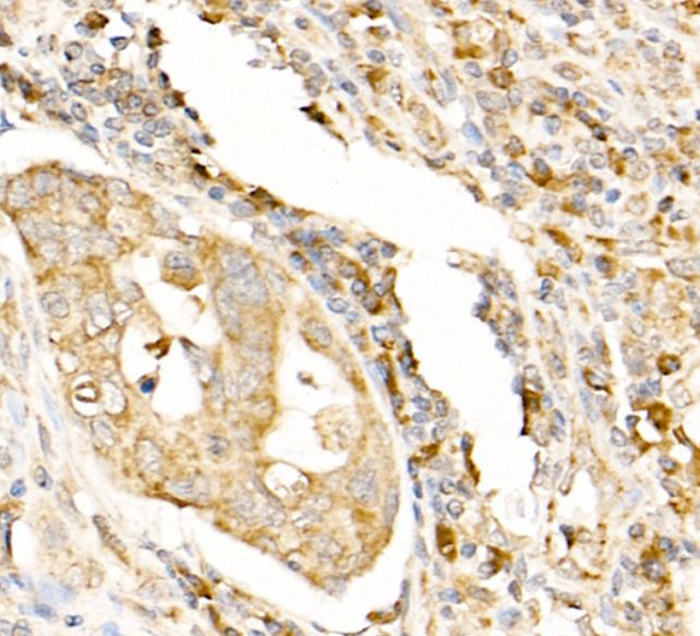
ARG46726 anti-LC3A/B antibody IHC-P image
Immunohistochemistry: Human colon carcinom stained with ARG46726 anti-LC3A/B antibody.
-

ARG46726 anti-LC3A/B antibody ICC/IF image
Immunofluorescence: 293T were treated with chloroquine and then stained with ARG46726 anti-LC3A/B antibody.
-
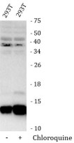
ARG46726 anti-LC3A/B antibody WB image
Western blot: 293T were treated with chloroquine and then stained with ARG46726 anti-LC3A/B antibody.
-

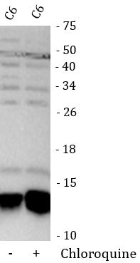
ARG46726 anti-LC3A/B antibody WB image
Western blot: C6 were treated with chloroquine and then stained with ARG46726 anti-LC3A/B antibody.
-
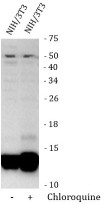
ARG46726 anti-LC3A/B antibody WB image
Western blot: NIH/3T3 were treated with chloroquine and then stained with ARG46726 anti-LC3A/B antibody.