ARG46262
anti-LRFN2 antibody
anti-LRFN2 antibody for Western blot,IHC-Formalin-fixed paraffin-embedded sections and Human,Mouse,Rat
Overview
| Product Description | Rabbit Polyclonal antibody recognizes LRFN2 |
|---|---|
| Tested Reactivity | Hu, Ms, Rat |
| Tested Application | IHC-P, WB |
| Host | Rabbit |
| Clonality | Polyclonal |
| Isotype | IgG |
| Target Name | LRFN2 |
| Antigen Species | Human |
| Immunogen | A 14 amino acid synthetic peptide within aa. 700 - 750 of human LRFN2. |
| Conjugation | Un-conjugated |
| Alternate Names | leucine rich repeat and fibronectin type III domain containing 2; LRFN2; SALM1; FIGLER2; KIAA1246; SALM1; Leucine-rich repeat and fibronectin type-III domain-containing protein 2; Synaptic adhesion-like molecule 1 |
Application Instructions
| Application Suggestion |
|
||||||
|---|---|---|---|---|---|---|---|
| Application Note | * The dilutions indicate recommended starting dilutions and the optimal dilutions or concentrations should be determined by the scientist. | ||||||
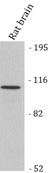
| Observed Size | 100 kDa |
Properties
| Form | Liquid |
|---|---|
| Purification | Affinity chromatography purified |
| Buffer | PBS and 0.02% Sodium azide. |
| Preservative | 0.02% Sodium azide |
| Concentration | 1 mg/ml |
| Storage Instruction | For continuous use, store undiluted antibody at 2-8°C for up to a week. For long-term storage, aliquot and store at -312°C or below. Storage in frost free freezers is not recommended. Avoid repeated freeze/thaw cycles. Suggest spin the vial prior to opening. The antibody solution should be gently mixed before use. |
| Note | For laboratory research only, not for drug, diagnostic or other use. |
Bioinformation
| Database Links |
Swiss-port # Q80TG9 Mouse Leucine-rich repeat and fibronectin type-III domain-containing protein 2 Swiss-port # Q9ULH4 Human Leucine-rich repeat and fibronectin type-III domain-containing protein 2 |
|---|---|
| Gene Symbol | LRFN2 |
| Gene Full Name | leucine rich repeat and fibronectin type III domain containing 2 |
| Background | Predicted to be involved in modulation of chemical synaptic transmission and regulation of postsynapse organization. Predicted to be located in plasma membrane. Predicted to be active in several cellular components, including Schaffer collateral - CA1 synapse; cell surface; and postsynaptic density membrane. [provided by Alliance of Genome Resources, Apr 2025] |
| Function | Promotes neurite outgrowth in hippocampal neurons. Enhances the cell surface expression of 2 NMDA receptor subunits GRIN1 and GRIN2A. May play a role in redistributing DLG4 to the cell periphery (By similarity). [UniProt] |
| Cellular Localization | Cell membrane. [UniProt] |
| Calculated MW | 90 kDa |
| PTM | Disulfide bond; Glycoprotein. [UniProt] |
Images (2) Click the Picture to Zoom In
-

ARG46262 anti-LRFN2 antibody WB image
Western blot: Rat brain stained with ARG46262 anti-LRFN2 antibody.
-

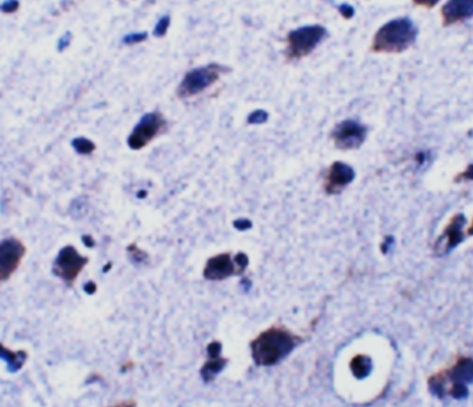
ARG46262 anti-LRFN2 antibody IHC-P image
Immunohistochemistry: Mouse brain stained with ARG46262 anti-LRFN2 antibody.