ARG45541
anti-LYPD3 antibody
anti-LYPD3 antibody for Flow cytometry,IHC-Formalin-fixed paraffin-embedded sections,Western blot and Human,Mouse,Rat
Overview
| Product Description | Rabbit Polyclonal antibody recognizes LYPD3 |
|---|---|
| Tested Reactivity | Hu, Ms, Rat |
| Tested Application | FACS, IHC-P, WB |
| Host | Rabbit |
| Clonality | Polyclonal |
| Isotype | IgG |
| Target Name | LYPD3 |
| Antigen Species | Human |
| Immunogen | Recombinant protein containing to human LYPD3. |
| Conjugation | Un-conjugated |
| Alternate Names | LYPD3; LY6/PLAUR domain containing 3; C4.4A; Ly6/PLAUR domain-containing protein 3; GPI-anchored metastasis-associated protein C4.4A homolog; MIG-C4; Matrigel-induced gene C4 protein |
Application Instructions
| Application Suggestion |
|
||||||||
|---|---|---|---|---|---|---|---|---|---|
| Application Note | * The dilutions indicate recommended starting dilutions and the optimal dilutions or concentrations should be determined by the scientist. | ||||||||
| Observed Size | 75 kDa |
Properties
| Form | Liquid |
|---|---|
| Purification | Affinity purified |
| Buffer | 0.2% Na2HPO4, 0.9% NaCl and 4% Trehalose. |
| Stabilizer | 4% Trehalose |
| Concentration | 0.5 mg/ml |
| Storage Instruction | For continuous use, store undiluted antibody at 2-8°C for up to a week. For long-term storage, aliquot and store at -20°C or below. Storage in frost free freezers is not recommended. Avoid repeated freeze/thaw cycles. Suggest spin the vial prior to opening. The antibody solution should be gently mixed before use. |
| Note | For laboratory research only, not for drug, diagnostic or other use. |
Bioinformation
| Database Links | |
|---|---|
| Gene Symbol | LYPD3 |
| Gene Full Name | LY6/PLAUR domain containing 3 |
| Background | Predicted to enable laminin binding activity. Involved in negative regulation of smooth muscle cell apoptotic process. Located in extracellular space. [provided by Alliance of Genome Resources, Feb 2025] |
| Function | Supports cell migration. May be involved in urothelial cell-matrix interactions. May be involved in tumor progression. [UniProt] |
| Cellular Localization | Cell membrane; Lipid-anchor, GPI-anchor. [UniProt] |
| Calculated MW | 36 kDa |
| PTM | N-glycosylated and O-glycosylated. [UniProt] |
Images (6) Click the Picture to Zoom In
-

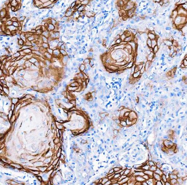
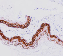
ARG45541 anti-LYPD3 antibody IHC-P image
Immunohistochemistry: Human skin cance stained with ARG45541 anti-LYPD3 antibody at 2 μg/ml dilution.
-
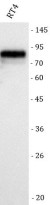
ARG45541 anti-LYPD3 antibody WB image
Western blot: RT4 stained with ARG45541 anti-LYPD3 antibody at 0.5 μg/ml dilution.
-
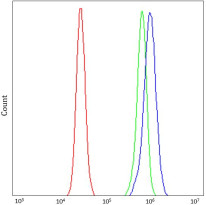
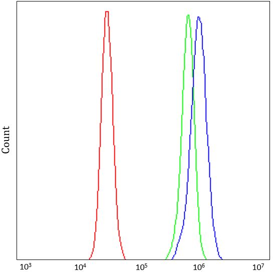
ARG45541 anti-LYPD3 antibody FACS image
Flow Cytometry: RT4 stained with ARG45541 anti-LYPD3 antibody at 1 µg/10^6 cells dilution.
-
ARG45541 anti-LYPD3 antibody IHC-P image
Immunohistochemistry: Rat stomach stained with ARG45541 anti-LYPD3 antibody at 2 μg/ml dilution.
-
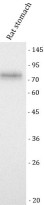
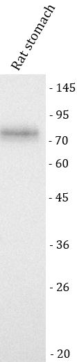
ARG45541 anti-LYPD3 antibody WB image
Western blot: Rat stomach stained with ARG45541 anti-LYPD3 antibody at 0.5 μg/ml dilution.
-
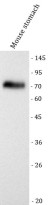
ARG45541 anti-LYPD3 antibody WB image
Western blot: Mouse stomach stained with ARG45541 anti-LYPD3 antibody at 0.5 μg/ml dilution.