ARG52326
anti-Lamin A + C antibody [4C4]
anti-Lamin A + C antibody [4C4] for ICC/IF,Western blot and Human,Mouse,Rat,Bovine
Controls and Markers antibody; Signaling Transduction antibody

Overview
| Product Description | Mouse Monoclonal antibody [4C4] recognizes Lamin A + C |
|---|---|
| Tested Reactivity | Hu, Ms, Rat, Bov |
| Tested Application | ICC/IF, WB |
| Host | Mouse |
| Clonality | Monoclonal |
| Clone | 4C4 |
| Isotype | IgG1 |
| Target Name | Lamin A + C |
| Antigen Species | Human |
| Immunogen | Recombinant full length human lamin C expressed in and purified from E. Coli. |
| Conjugation | Un-conjugated |
| Alternate Names | HGPS; Renal carcinoma antigen NY-REN-32; LDP1; FPL; LMN1; CDCD1; LMNL1; CDDC; PRO1; EMD2; CMT2B1; 70 kDa lamin; LFP; Prelamin-A/C; LMNC; FPLD2; LGMD1B; IDC; FPLD; CMD1A |
Application Instructions
| Application Suggestion |
|
||||||
|---|---|---|---|---|---|---|---|
| Application Note | Specific for the ~64 and 74k lamin A and C proteins. * The dilutions indicate recommended starting dilutions and the optimal dilutions or concentrations should be determined by the scientist. |
Properties
| Form | Liquid |
|---|---|
| Purification | Affinity Purified |
| Buffer | PBS and 10 mM Sodium azide |
| Preservative | 10 mM Sodium azide |
| Storage Instruction | For continuous use, store undiluted antibody at 2-8°C for up to a week. For long-term storage, aliquot and store at -20°C or below. Storage in frost free freezers is not recommended. Avoid repeated freeze/thaw cycles. Suggest spin the vial prior to opening. The antibody solution should be gently mixed before use. |
| Note | For laboratory research only, not for drug, diagnostic or other use. |
Bioinformation
| Database Links | |
|---|---|
| Gene Symbol | LMNA |
| Gene Full Name | lamin A/C |
| Background | Lamins A and C are nuclear structural proteins that are part of the intermediate filament family and coded for by the same gene (LMNA). Lamins A and C are nearly identical except for their carboxy termini (McKeon et al., 1986). Mutations in the gene encoding lamins A/C have been shown to cause a variety of diseases including autosomal dominant Emery-Dreifuss muscular dystrophy (Bonne et al., 1995), autosomal dominant limbgirdle muscular dystrophy (Muchir et al., 2000) and Charcot-Marie-Tooth disorder type 2 (De Sandre-Giavonnoli et al., 2002). |
| Research Area | Controls and Markers antibody; Signaling Transduction antibody |
| Calculated MW | Lamin A: 74 kDa Lamin C: 65 kDa |
| PTM | Increased phosphorylation of the lamins occurs before envelope disintegration and probably plays a role in regulating lamin associations. Proteolytic cleavage of the C-terminal of 18 residues of prelamin-A/C results in the production of lamin-A/C. The prelamin-A/C maturation pathway includes farnesylation of CAAX motif, ZMPSTE24/FACE1 mediated cleavage of the last three amino acids, methylation of the C-terminal cysteine and endoproteolytic removal of the last 15 C-terminal amino acids. Proteolytic cleavage requires prior farnesylation and methylation, and absence of these blocks cleavage. Sumoylation is necessary for the localization to the nuclear envelope. Farnesylation of prelamin-A/C facilitates nuclear envelope targeting. |
Images (5) Click the Picture to Zoom In
-
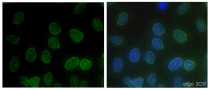
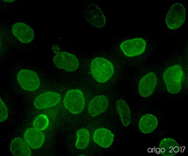
ARG52326 anti-Lamin A + C antibody [4C4] ICC/IF image
Immunofluorescence: 100% Methanol fixed (RT, 10 min) HeLa cells stained with ARG52326 anti-Lamin A + C antibody [4C4] at 1:100 dilution. Left: primary antibody (green). Right: Merge (primary antibody and DAPI).
Secondary antibody: ARG55393 Goat anti-Mouse IgG (H+L) antibody (FITC)
-
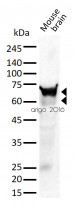
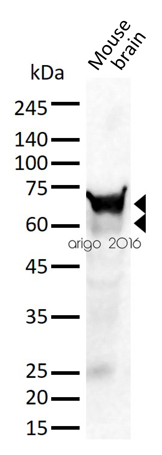
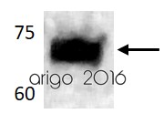
ARG52326 anti-Lamin A + C antibody [4C4] WB image
Western blot: 30 µg of Mouse brain lysate stained with ARG52326 anti-Lamin A + C antibody [4C4] at 1:1000 dilution.
-

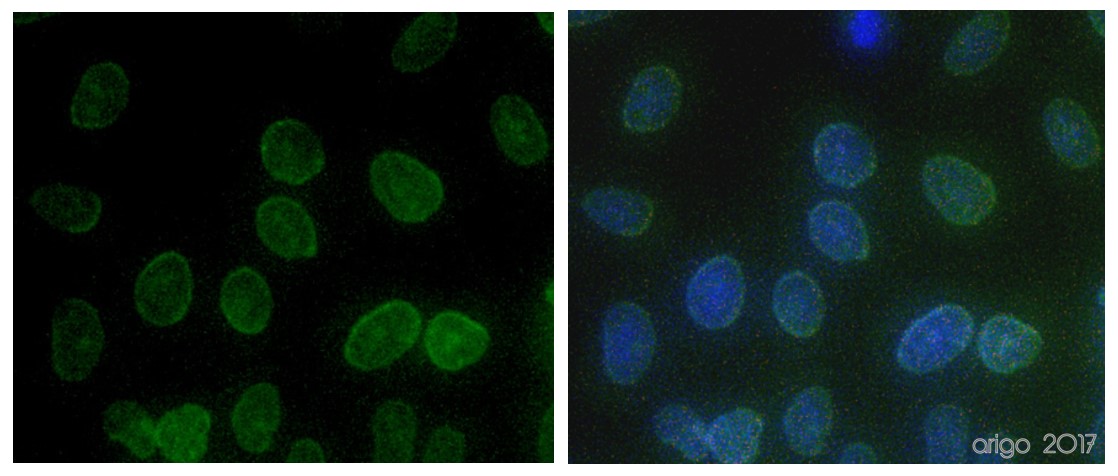
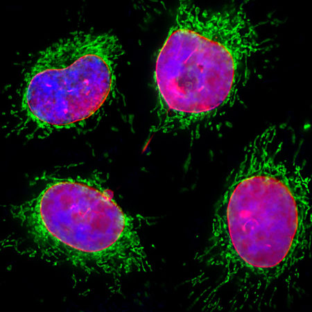
ARG52326 anti-Lamin A + C antibody [4C4] ICC/IF image
Immunofluorescence: HeLa cells stained with ARG52326 anti-Lamin A + C antibody [4C4] (red) at 1:2000 dilution, and costained with anti-Hsp 60 antibody (green) at 1:5000 dilution. Hoechst (blue) for nuclear staining.
Clone 4C4 specifically labels the nuclear lamina, while Hsp 60 antibody reveals protein expressed in mitochondria.
-
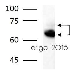
ARG52326 anti-Lamin A + C antibody [4C4] WB image
Western blot: HeLa lysate showing specific immunolabeling of the ~ 64k and 74k lamin A/C proteins stained with ARG52326 anti-Lamin A + C antibody [4C4].
-

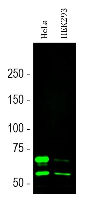
ARG52326 anti-Lamin A + C antibody [4C4] WB image
Western blot: HeLa and HEK293 cell lysates stained with ARG52326 anti-Lamin A + C antibody [4C4] (green) at 1:1000 dilution.
Two strong bands at ~74 and 65 kDa correspond to the lamin A and lamin C proteins respectively.
Customer's Feedback
Excellent
Review for anti-Lamin A + C antibody [4C4]
Application:IF/ICC
Sample:HeLa
Fixation Buffer:100% Methanol
Fixation Time:10 min
Fixation Temperature:RT ºC
Permeabilization Buffer:0.1% Triton X-100
Primary Antibody Dilution Factor:1:100
Primary Antibody Incubation Time:overnight
Primary Antibody Incubation Temperature:4 ºC
Conjugation of Secondary Antibody:FITC

Excellent
Review for anti-Lamin A + C antibody [4C4]
Application:WB
Sample:Rat brain
Sample Loading Amount:30 µg
Primary Antibody Dilution Factor:1:1000
Primary Antibody Incubation Time:overnight
Primary Antibody Incubation Temperature:4 ºC
Excellent
Review for anti-Lamin A + C antibody [4C4]
Application:WB
Sample:PC-3
Sample Loading Amount:30 µg
Primary Antibody Dilution Factor:1:1000
Primary Antibody Incubation Time:overnight
Primary Antibody Incubation Temperature:4 ºC


