ARG67322
anti-MHC Class II beta antibody
anti-MHC Class II beta antibody for Western blot,IHC-Formalin-fixed paraffin-embedded sections,ICC/IF and Human,Mouse,Rat
Overview
| Product Description | Rabbit Monoclonal antibody recognizes MHC Class II beta |
|---|---|
| Tested Reactivity | Hu, Ms, Rat |
| Tested Application | ICC/IF, IHC-P, WB |
| Host | Rabbit |
| Clonality | Monoclonal |
| Target Name | MHC Class II beta |
| Antigen Species | Human |
| Immunogen | KLH-conjugated synthetic peptide encompassing a sequence within encompassing a sequence within human MHC Class II beta |
| Conjugation | Un-conjugated |
| Alternate Names | HLA-DP1B; HLA class II histocompatibility antigen, DP beta 1 chain; HLA class II histocompatibility antigen, DP(W4) beta chain; MHC class II antigen DPB1 |
Application Instructions
| Application Suggestion |
|
||||||||
|---|---|---|---|---|---|---|---|---|---|
| Application Note | * The dilutions indicate recommended starting dilutions and the optimal dilutions or concentrations should be determined by the scientist. | ||||||||
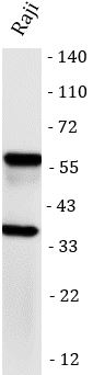
| Observed Size | 25-35 kDa(Monomer); 50-65 kDa(Dimer) |
Properties
| Form | Liquid |
|---|---|
| Purification | Affinity purification with immunogen? |
| Buffer | PBS, 0.05% Proclin300. 50% Glycerol, and 0.5% BSA |
| Preservative | 0.01% Sodium azide |
| Stabilizer | 50% Glycerol, and 0.5% BSA |
| Storage Instruction | For continuous use, store undiluted antibody at 2-8°C for up to a week. For long-term storage, aliquot and store at -20°C or below. Storage in frost free freezers is not recommended. Avoid repeated freeze/thaw cycles. Suggest spin the vial prior to opening. The antibody solution should be gently mixed before use. |
| Note | For laboratory research only, not for drug, diagnostic or other use. |
Bioinformation
| Database Links |
Swiss-port # P04440 Human HLA class II histocompatibility antigen, DP beta 1 chain |
|---|---|
| Gene Symbol | HLA-DPB1 |
| Gene Full Name | Major Histocompatibility Complex, Class II, DP Beta 1 |
| Background | HLA-DPB belongs to the HLA class II beta chain paralogues. This class II molecule is a heterodimer consisting of an alpha (DPA) and a beta chain (DPB), both anchored in the membrane. It plays a central role in the immune system by presenting peptides derived from extracellular proteins. Class II molecules are expressed in antigen presenting cells (APC: B lymphocytes, dendritic cells, macrophages). The beta chain is approximately 26-28 kDa and its gene contains 6 exons. Exon one encodes the leader peptide, exons 2 and 3 encode the two extracellular domains, exon 4 encodes the transmembrane domain and exon 5 encodes the cytoplasmic tail. Within the DP molecule both the alpha chain and the beta chain contain the polymorphisms specifying the peptide binding specificities, resulting in up to 4 different molecules. [provided by RefSeq, Jul 2008] |
| Function | Binds peptides derived from antigens that access the endocytic route of antigen presenting cells (APC) and presents them on the cell surface for recognition by the CD4 T-cells. The peptide binding cleft accommodates peptides of 10-30 residues. The peptides presented by MHC class II molecules are generated mostly by degradation of proteins that access the endocytic route, where they are processed by lysosomal proteases and other hydrolases. Exogenous antigens that have been endocytosed by the APC are thus readily available for presentation via MHC II molecules, and for this reason this antigen presentation pathway is usually referred to as exogenous. As membrane proteins on their way to degradation in lysosomes as part of their normal turn-over are also contained in the endosomal/lysosomal compartments, exogenous antigens must compete with those derived from endogenous components. Autophagy is also a source of endogenous peptides, autophagosomes constitutively fuse with MHC class II loading compartments. In addition to APCs, other cells of the gastrointestinal tract, such as epithelial cells, express MHC class II molecules and CD74 and act as APCs, which is an unusual trait of the GI tract. To produce a MHC class II molecule that presents an antigen, three MHC class II molecules (heterodimers of an alpha and a beta chain) associate with a CD74 trimer in the ER to form a heterononamer. Soon after the entry of this complex into the endosomal/lysosomal system where antigen processing occurs, CD74 undergoes a sequential degradation by various proteases, including CTSS and CTSL, leaving a small fragment termed CLIP (class-II-associated invariant chain peptide). The removal of CLIP is facilitated by HLA-DM via direct binding to the alpha-beta-CLIP complex so that CLIP is released. HLA-DM stabilizes MHC class II molecules until primary high affinity antigenic peptides are bound. The MHC II molecule bound to a peptide is then transported to the cell membrane surface. In B-cells, the interaction between HLA-DM and MHC class II molecules is regulated by HLA-DO. Primary dendritic cells (DCs) also to express HLA-DO. Lysosomal microenvironment has been implicated in the regulation of antigen loading into MHC II molecules, increased acidification produces increased proteolysis and efficient peptide loading. [UniProt] |
| Cellular Localization | Cell membrane; Endoplasmic reticulum. [UniProt] |
| Calculated MW | 29 kDa |
| PTM | Disulfide bond; Glycoprotein. [UniProt] |
Images (3) Click the Picture to Zoom In
-
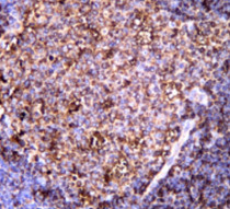
ARG67322 anti-MHC Class II beta antibody IHC-P image
Immunohistochemistry: Human tonsil stained with ARG67322 anti-MHC Class II beta antibody.
-
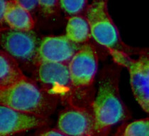
ARG67322 anti-MHC Class II beta antibody ICC/IF image
Immunofluorescence: Raji stained with ARG67322 anti-MHC Class II beta antibody.
-

ARG67322 anti-MHC Class II beta antibody WB image
Western blot: Raji stained with ARG67322 anti-MHC Class II beta antibody.