ARG45467
anti-NEDD9 antibody
anti-NEDD9 antibody for Flow cytometry,ICC/IF,IHC-Formalin-fixed paraffin-embedded sections,Western blot and Human,Mouse,Rat,Monkey
Overview
| Product Description | Rabbit Polyclonal antibody recognizes NEDD9 |
|---|---|
| Tested Reactivity | Hu, Ms, Rat, Mk |
| Tested Application | FACS, ICC/IF, IHC-P, WB |
| Host | Rabbit |
| Clonality | Polyclonal |
| Isotype | IgG |
| Target Name | NEDD9 |
| Antigen Species | Human |
| Immunogen | Recombinant protein containing to human NEDD9. |
| Conjugation | Un-conjugated |
| Alternate Names | CASS2; Renal carcinoma antigen NY-REN-12; hEF1; HEF1; p105; CASL; CAS-L; Cas scaffolding protein family member 2; CRK-associated substrate-related protein; CasL; Enhancer of filamentation 1; CAS2; Neural precursor cell expressed developmentally down-regulated protein 9; NEDD-9 |
Application Instructions
| Application Suggestion |
|
||||||||||
|---|---|---|---|---|---|---|---|---|---|---|---|
| Application Note | * The dilutions indicate recommended starting dilutions and the optimal dilutions or concentrations should be determined by the scientist. | ||||||||||
| Observed Size | 93 - 105 kDa |
Properties
| Form | Liquid |
|---|---|
| Purification | Affinity purified |
| Buffer | 0.2% Na2HPO4, 0.9% NaCl and 4% Trehalose. |
| Stabilizer | 4% Trehalose |
| Concentration | 0.5 mg/ml |
| Storage Instruction | For continuous use, store undiluted antibody at 2-8°C for up to a week. For long-term storage, aliquot and store at -20°C or below. Storage in frost free freezers is not recommended. Avoid repeated freeze/thaw cycles. Suggest spin the vial prior to opening. The antibody solution should be gently mixed before use. |
| Note | For laboratory research only, not for drug, diagnostic or other use. |
Bioinformation
| Database Links | |
|---|---|
| Gene Symbol | NEDD9 |
| Gene Full Name | Neural Precursor Cell Expressed, Developmentally Down-Regulated 9 |
| Background | The protein encoded by this gene is a member of the CRK-associated substrates family. Members of this family are adhesion docking molecules that mediate protein-protein interactions for signal transduction pathways. This protein is a focal adhesion protein that acts as a scaffold to regulate signaling complexes important in cell attachment, migration and invasion as well as apoptosis and the cell cycle. This protein has also been reported to have a role in cancer metastasis. Alternative splicing results in multiple transcript variants. [provided by RefSeq, Aug 2012] |
| Function | Scaffolding protein which plays a central coordinating role for tyrosine-kinase-based signaling related to cell adhesion [UniProt] |
| Cellular Localization | Cell junction; Cell membrane; Cell projection; Cytoplasm; Cytoskeleton; Golgi apparatus; Membrane; Nucleus. [UniProt] |
| Calculated MW | 93 kDa |
| PTM | Cell cycle-regulated processing produces four isoforms: p115, p105, p65, and p55. Isoform p115 arises from p105 phosphorylation and appears later in the cell cycle. Isoform p55 arises from p105 as a result of cleavage at a caspase cleavage-related site and it appears specifically at mitosis. The p65 isoform is poorly detected. PTK2/FAK1 phosphorylates the protein at the YDYVHL motif (conserved among all cas proteins). The SRC family kinases (FYN, SRC, LCK and CRK) are recruited to the phosphorylated sites and can phosphorylate other tyrosine residues. Ligation of either integrin beta-1 or B-cell antigen receptor on tonsillar B-cells and B-cell lines promotes tyrosine phosphorylation and both integrin and BCR-mediated tyrosine phosphorylation requires an intact actin network. In fibroblasts transformation with oncogene v-ABL results in an increase in tyrosine phosphorylation. Transiently phosphorylated following CD3 cross-linking and this phosphorylated form binds to CRK and C3G. A mutant lacking the SH3 domain is phosphorylated upon CD3 cross-linking but not upon integrin beta-1 cross-linking. Tyrosine phosphorylation occurs upon stimulation of the G-protein coupled C1a calcitonin receptor. Calcitonin-stimulated tyrosine phosphorylation is mediated by calcium- and protein kinase C-dependent mechanisms and requires the integrity of the actin cytoskeleton. Phosphorylation at Ser-369 induces proteasomal degradation. [UniProt] |
Images (7) Click the Picture to Zoom In
-

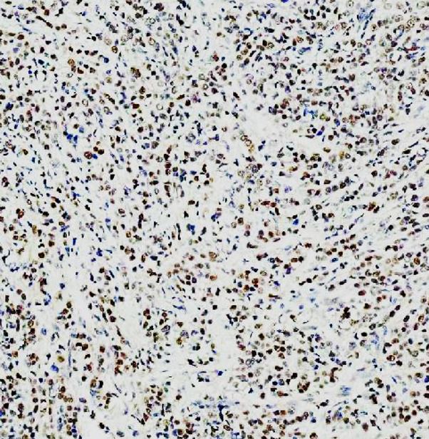
ARG45467 anti-NEDD9 antibody IHC-P image
Immunohistochemistry: Human breast cancer stained with ARG45467 anti-NEDD9 antibody at 2 μg/ml dilution.
-

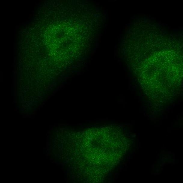
ARG45467 anti-NEDD9 antibody ICC/IF image
Immunofluorescence: Hela stained with ARG45467 anti-NEDD9 antibody at 5 μg/ml dilution.
-
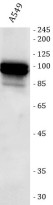
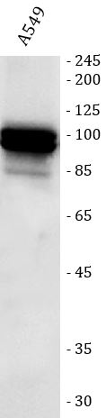
ARG45467 anti-NEDD9 antibody WB image
Western blot: A549 stained with ARG45467 anti-NEDD9 antibody at 0.5 μg/ml dilution.
-

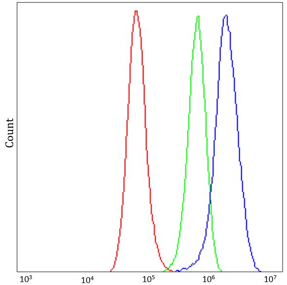
ARG45467 anti-NEDD9 antibody FACS image
Flow Cytometry: A549 stained with ARG45467 anti-NEDD9 antibody at 1 µg/10^6 cells dilution.
-
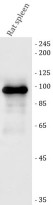
ARG45467 anti-NEDD9 antibody WB image
Western blot: Rat spleen stained with ARG45467 anti-NEDD9 antibody at 0.5 μg/ml dilution.
-
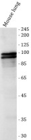
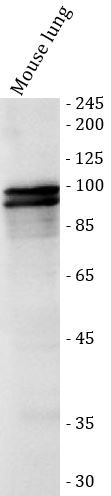
ARG45467 anti-NEDD9 antibody WB image
Western blot: Mouse lung stained with ARG45467 anti-NEDD9 antibody at 0.5 μg/ml dilution.
-

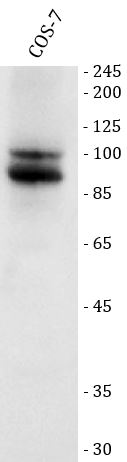
ARG45467 anti-NEDD9 antibody WB image
Western blot: COS-7 stained with ARG45467 anti-NEDD9 antibody at 0.5 μg/ml dilution.