ARG40514
anti-OAT antibody
anti-OAT antibody for Flow cytometry,ICC/IF,IHC-Formalin-fixed paraffin-embedded sections,Western blot and Human,Mouse,Rat
Overview
| Product Description | Rabbit Polyclonal antibody recognizes OAT |
|---|---|
| Tested Reactivity | Hu, Ms, Rat |
| Tested Application | FACS, ICC/IF, IHC-P, WB |
| Host | Rabbit |
| Clonality | Polyclonal |
| Isotype | IgG |
| Target Name | OAT |
| Antigen Species | Human |
| Immunogen | KLH-conjugated synthetic peptide corresponding to aa. 27-55 of Human OAT. |
| Conjugation | Un-conjugated |
| Alternate Names | OATASE; OKT; Ornithine aminotransferase, mitochondrial; Ornithine--oxo-acid aminotransferase; GACR; HOGA; Ornithine delta-aminotransferase; EC 2.6.1.13 |
Application Instructions
| Application Suggestion |
|
||||||||||
|---|---|---|---|---|---|---|---|---|---|---|---|
| Application Note | * The dilutions indicate recommended starting dilutions and the optimal dilutions or concentrations should be determined by the scientist. | ||||||||||
| Positive Control | 293 |
Properties
| Form | Liquid |
|---|---|
| Purification | Purification with Protein A and immunogen peptide. |
| Buffer | PBS and 0.09% (W/V) Sodium azide. |
| Preservative | 0.09% (W/V) Sodium azide. |
| Storage Instruction | For continuous use, store undiluted antibody at 2-8°C for up to a week. For long-term storage, aliquot and store at -20°C or below. Storage in frost free freezers is not recommended. Avoid repeated freeze/thaw cycles. Suggest spin the vial prior to opening. The antibody solution should be gently mixed before use. |
| Note | For laboratory research only, not for drug, diagnostic or other use. |
Bioinformation
| Database Links | |
|---|---|
| Gene Symbol | OAT |
| Gene Full Name | ornithine aminotransferase |
| Background | This gene encodes the mitochondrial enzyme ornithine aminotransferase, which is a key enzyme in the pathway that converts arginine and ornithine into the major excitatory and inhibitory neurotransmitters glutamate and GABA. Mutations that result in a deficiency of this enzyme cause the autosomal recessive eye disease Gyrate Atrophy. Alternatively spliced transcript variants encoding different isoforms have been described. Related pseudogenes have been defined on the X chromosome. [provided by RefSeq, Jan 2010] |
| Cellular Localization | Mitochondrion matrix. [UniProt] |
| Calculated MW | 49 kDa |
Images (4) Click the Picture to Zoom In
-
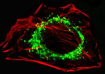
ARG40514 anti-OAT antibody ICC/IF image
Immunofluorescence: A549 cells were fixed with 4% PFA (20 min), permeabilized with Triton X-100 (0.1%, 10 min), then stained with ARG40514 anti-OAT antibody (green) at 1:25, 1 hour at 37°C. Cytoplasmic actin was counterstained with Alexa Fluor® 555 conjugated Phalloidin (red).
-
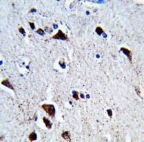
ARG40514 anti-OAT antibody IHC-P image
Immunohistochemistry: Formalin-fixed and paraffin-embedded Human brain tissue stained with ARG40514 anti-OAT antibody.
-
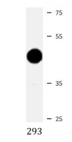
ARG40514 anti-OAT antibody WB image
Western blot: 20 µg of 293 whole cell lysate stained with ARG40514 anti-OAT antibody at 1:1000 dilution.
-
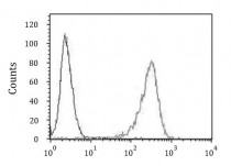
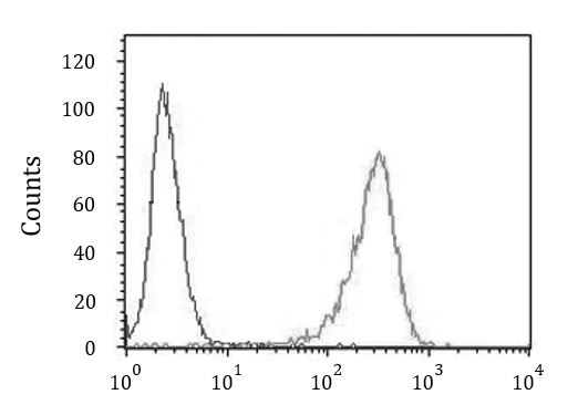
ARG40514 anti-OAT antibody FACS image
Flow Cytometry: 293 cells stained with ARG40514 anti-OAT antibody (right histogram) or without primary antibody as control (left histogram), followed by incubation with FITC labelled secondary antibody.