ARG59224
anti-PTP4A2 antibody
anti-PTP4A2 antibody for Flow cytometry,IHC-Formalin-fixed paraffin-embedded sections,Western blot and Human,Mouse,Rat
Overview
| Product Description | Rabbit Polyclonal antibody recognizes PTP4A2 |
|---|---|
| Tested Reactivity | Hu, Ms, Rat |
| Predict Reactivity | Hm |
| Tested Application | FACS, IHC-P, WB |
| Host | Rabbit |
| Clonality | Polyclonal |
| Isotype | IgG |
| Target Name | PTP4A2 |
| Antigen Species | Human |
| Immunogen | Synthetic peptide corresponding to aa. 40-69 of Human PTP4A2. (TTLVRVCDATYDKAPVEKEGIHVLDWPFDD) |
| Conjugation | Un-conjugated |
| Alternate Names | OV-1; Protein-tyrosine phosphatase of regenerating liver 2; PTP4A; Protein tyrosine phosphatase type IVA 2; PTPCAAX2; PRL2; ptp-IV1b; ptp-IV1a; CAAXII; PRL-2; HH13; Protein-tyrosine phosphatase 4a2; EC 3.1.3.48; HU-PP-1; PTP; HH7-2 |
Application Instructions
| Application Suggestion |
|
||||||||
|---|---|---|---|---|---|---|---|---|---|
| Application Note | IHC-P: Antigen Retrieval: Heat mediated was performed in Citrate buffer (pH 6.0) for 20 min. * The dilutions indicate recommended starting dilutions and the optimal dilutions or concentrations should be determined by the scientist. |
Properties
| Form | Liquid |
|---|---|
| Purification | Affinity purification with immunogen. |
| Buffer | 0.9% NaCl, 0.2% Na2HPO4, 0.05% Sodium azide and 5% BSA. |
| Preservative | 0.05% Sodium azide |
| Stabilizer | 5% BSA |
| Concentration | 0.5 mg/ml |
| Storage Instruction | For continuous use, store undiluted antibody at 2-8°C for up to a week. For long-term storage, aliquot and store at -20°C or below. Storage in frost free freezers is not recommended. Avoid repeated freeze/thaw cycles. Suggest spin the vial prior to opening. The antibody solution should be gently mixed before use. |
| Note | For laboratory research only, not for drug, diagnostic or other use. |
Bioinformation
| Database Links | |
|---|---|
| Gene Symbol | PTP4A2 |
| Gene Full Name | protein tyrosine phosphatase type IVA, member 2 |
| Background | The protein encoded by this gene belongs to a small class of the protein tyrosine phosphatase (PTP) family. PTPs are cell signaling molecules that play regulatory roles in a variety of cellular processes. PTPs in this class contain a protein tyrosine phosphatase catalytic domain and a characteristic C-terminal prenylation motif. This PTP has been shown to primarily associate with plasmic and endosomal membrane through its C-terminal prenylation. This PTP was found to interact with the beta-subunit of Rab geranylgeranyltransferase II (beta GGT II), and thus may function as a regulator of GGT II activity. Overexpression of this gene in mammalian cells conferred a transformed phenotype, which suggested its role in tumorigenesis. Alternatively spliced transcript variants have been described. Related pseudogenes exist on chromosomes 11, 12 and 17. [provided by RefSeq, Aug 2010] |
| Function | Protein tyrosine phosphatase which stimulates progression from G1 into S phase during mitosis. Promotes tumors. Inhibits geranylgeranyl transferase type II activity by blocking the association between RABGGTA and RABGGTB. [UniProt] |
| Cellular Localization | Cell membrane. Early endosome. Cytoplasm. [UniProt] |
| Calculated MW | 19 kDa |
| PTM | Farnesylated. Farnesylation is required for membrane targeting and for interaction with RABGGTB. Unfarnesylated forms are redirected to the nucleus and cytosol. [UniProt] |
Images (5) Click the Picture to Zoom In
-
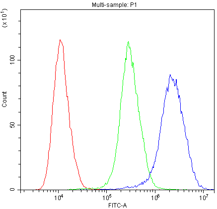
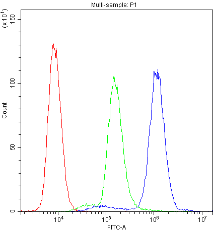
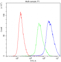
ARG59224 anti-PTP4A2 antibody FACS image
Flow Cytometry: U937 cells were blocked with 10% normal goat serum and then stained with ARG59224 anti-PTP4A2 antibody (blue) at 1 ug/10^6 cells for 30 min at 20°C, followed by DyLight®488 labelled secondary antibody. Isotype control antibody (green) was rabbit IgG (1 ug/10^6 cells) used under the same conditions. Unlabelled sample (red) was also used as a control.
-
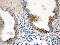
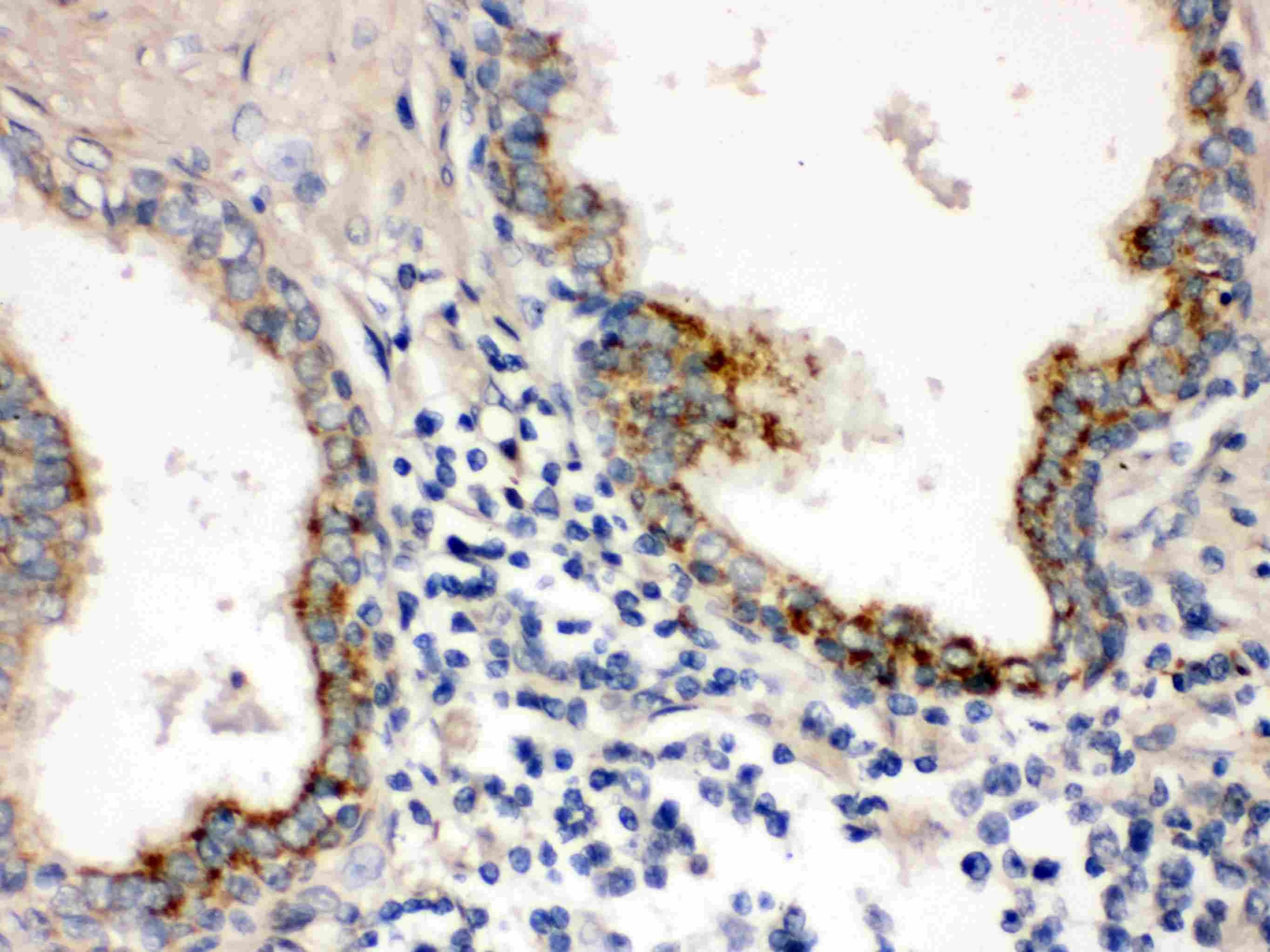
ARG59224 anti-PTP4A2 antibody IHC-P image
Immunohistochemistry: Paraffin-embedded Human prostatic cancer Tissue. Antigen Retrieval: Heat mediated was performed in Citrate buffer (pH 6.0, epitope retrieval solution) for 20 min. The tissue section was blocked with 10% goat serum. The tissue section was then stained with ARG59224 anti-PTP4A2 antibody at 1 µg/ml dilution, overnight at 4°C.
-
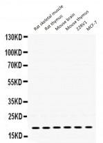
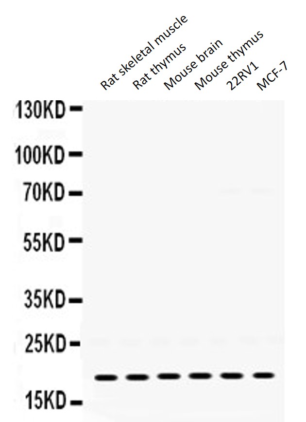
ARG59224 anti-PTP4A2 antibody WB image
Western blot: 50 µg of samples under reducing conditions. Rat skeletal muscle, Rat thymus, Mouse brain, Mouse thymus, 22RV1 and MCF-7 whole cell lysates stained with ARG59224 anti-PTP4A2 antibody at 0.5 µg/ml, overnight at 4°C.
-
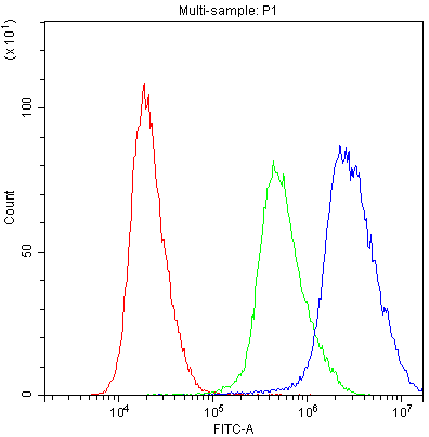
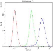
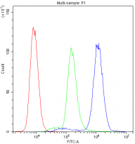
ARG59224 anti-PTP4A2 antibody FACS image
Flow Cytometry: PC-3 cells were blocked with 10% normal goat serum and then stained with ARG59224 anti-PTP4A2 antibody (blue) at 1 ug/10^6 cells for 30 min at 20°C, followed by DyLight®488 labelled secondary antibody. Isotype control antibody (green) was rabbit IgG (1 ug/10^6 cells) used under the same conditions. Unlabelled sample (red) was also used as a control.
-
ARG59224 anti-PTP4A2 antibody FACS image
Flow Cytometry: A431 cells were blocked with 10% normal goat serum and then stained with ARG59224 anti-PTP4A2 antibody (blue) at 1 ug/10^6 cells for 30 min at 20°C, followed by DyLight®488 labelled secondary antibody. Isotype control antibody (green) was rabbit IgG (1 ug/10^6 cells) used under the same conditions. Unlabelled sample (red) was also used as a control.