ARG45596
anti-T Plastin / PLS3 antibody
anti-T Plastin / PLS3 antibody for Flow cytometry,ICC/IF,Immunoprecipitation,Western blot and Human,Mouse,Rat
Overview
| Product Description | Rabbit Polyclonal antibody recognizes T Plastin / PLS3 |
|---|---|
| Tested Reactivity | Hu, Ms, Rat |
| Tested Application | FACS, ICC/IF, IP, WB |
| Host | Rabbit |
| Clonality | Polyclonal |
| Isotype | IgG |
| Target Name | T Plastin / PLS3 |
| Antigen Species | Human |
| Immunogen | Recombinant protein containing to human T Plastin / PLS3. |
| Conjugation | Un-conjugated |
| Alternate Names | PLS3; Plastin 3; T-Plastin; Plastin-3; Plastin 3 (T Isoform); T Fimbrin; T Plastin; T-Fimbrin; BMND18; DIH5 |
Application Instructions
| Application Suggestion |
|
||||||||||
|---|---|---|---|---|---|---|---|---|---|---|---|
| Application Note | * The dilutions indicate recommended starting dilutions and the optimal dilutions or concentrations should be determined by the scientist. | ||||||||||
| Observed Size | 71 kDa |
Properties
| Form | Liquid |
|---|---|
| Purification | Affinity purified |
| Buffer | 0.2% Na2HPO4, 0.9% NaCl and 4% Trehalose. |
| Stabilizer | 4% Trehalose |
| Concentration | 0.5 mg/ml |
| Storage Instruction | For continuous use, store undiluted antibody at 2-8°C for up to a week. For long-term storage, aliquot and store at -20°C or below. Storage in frost free freezers is not recommended. Avoid repeated freeze/thaw cycles. Suggest spin the vial prior to opening. The antibody solution should be gently mixed before use. |
| Note | For laboratory research only, not for drug, diagnostic or other use. |
Bioinformation
| Database Links | |
|---|---|
| Gene Symbol | PLS3 |
| Gene Full Name | Plastin 3 |
| Background | Plastins are a family of actin-binding proteins that are conserved throughout eukaryote evolution and expressed in most tissues of higher eukaryotes. In humans, two ubiquitous plastin isoforms (L and T) have been identified. Plastin 1 (otherwise known as Fimbrin) is a third distinct plastin isoform which is specifically expressed at high levels in the small intestine. The L isoform is expressed only in hemopoietic cell lineages, while the T isoform has been found in all other normal cells of solid tissues that have replicative potential (fibroblasts, endothelial cells, epithelial cells, melanocytes, etc.). The C-terminal 570 amino acids of the T-plastin and L-plastin proteins are 83% identical. It contains a potential calcium-binding site near the N terminus. Alternate splicing results in multiple transcript variants.[provided by RefSeq, Feb 2010] |
| Function | Actin-bundling protein. [UniProt] |
| Cellular Localization | Cytoplasm; Nucleus. [UniProt] |
| Calculated MW | 71 kDa |
| PTM | Phosphoprotein. [UniProt] |
Images (6) Click the Picture to Zoom In
-
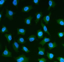
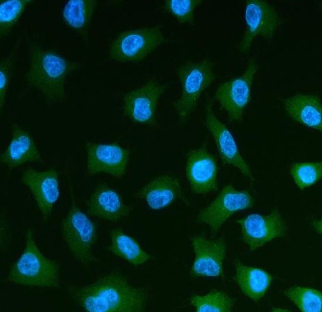
ARG45596 anti-T Plastin / PLS3 antibody ICC/IF image
Immunofluorescence: A549 stained with ARG45596 anti-T Plastin / PLS3 antibody at 5 μg/ml dilution.
-
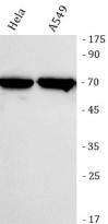
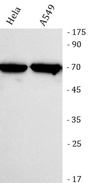
ARG45596 anti-T Plastin / PLS3 antibody WB image
Western blot: Hela and A549 stained with ARG45596 anti-T Plastin / PLS3 antibody at 0.5 μg/ml dilution.
-
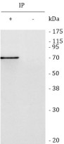
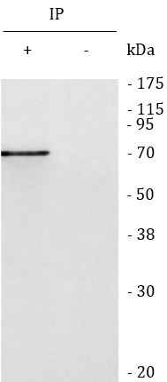
ARG45596 anti-T Plastin / PLS3 antibody IP image
Immunoprecipitation: Hela lysate immunoprecipitated with 2 μg ARG45596 anti-T Plastin / PLS3 antibody.
-
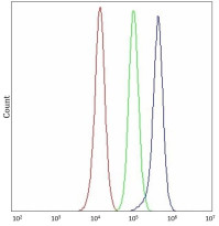
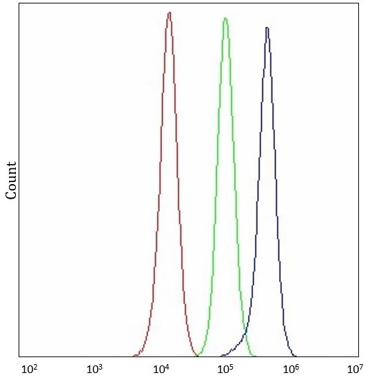
ARG45596 anti-T Plastin / PLS3 antibody FACS image
Flow Cytometry: PC-3 stained with ARG45596 anti-T Plastin / PLS3 antibody at 1 µg/10^6 cells dilution.
-

ARG45596 anti-T Plastin / PLS3 antibody WB image
Western blot: Rat stomach stained with ARG45596 anti-T Plastin / PLS3 antibody at 0.5 μg/ml dilution.
-

ARG45596 anti-T Plastin / PLS3 antibody WB image
Western blot: Mouse stomach stained with ARG45596 anti-T Plastin / PLS3 antibody at 0.5 μg/ml dilution.