ARG45186
anti-VDAC3 antibody
anti-VDAC3 antibody for Flow cytometry,IHC-Formalin-fixed paraffin-embedded sections,Western blot and Human,Mouse,Rat
Overview
| Product Description | Rabbit Polyclonal antibody recognizes VDAC3 |
|---|---|
| Tested Reactivity | Hu, Ms, Rat |
| Tested Application | FACS, IHC-P, WB |
| Host | Rabbit |
| Clonality | Polyclonal |
| Isotype | IgG |
| Target Name | VDAC3 |
| Antigen Species | Human |
| Immunogen | Recombinant protein containing to human VDAC3. |
| Conjugation | Un-conjugated |
| Alternate Names | VDAC3; Voltage Dependent Anion Channel 3; HD-VDAC3; Voltage-Dependent Anion-Selective Channel Protein 3; Outer Mitochondrial Membrane Protein Porin 3; VDAC-3; HVDAC3 |
Application Instructions
| Application Suggestion |
|
||||||||
|---|---|---|---|---|---|---|---|---|---|
| Application Note | * The dilutions indicate recommended starting dilutions and the optimal dilutions or concentrations should be determined by the scientist. | ||||||||
| Observed Size | 31 kDa |
Properties
| Form | Liquid |
|---|---|
| Purification | Affinity purification with immunogen. |
| Buffer | 0.9% NaCl, 0.2% Na2HPO4, 0.01% Sodium azide and 4% Trehalose. |
| Preservative | 0.01% Sodium azide |
| Stabilizer | 4% Trehalose |
| Concentration | 0.5 mg/ml |
| Storage Instruction | For continuous use, store undiluted antibody at 2-8°C for up to a week. For long-term storage, aliquot and store at -20°C or below. Storage in frost free freezers is not recommended. Avoid repeated freeze/thaw cycles. Suggest spin the vial prior to opening. The antibody solution should be gently mixed before use. |
| Note | For laboratory research only, not for drug, diagnostic or other use. |
Bioinformation
| Database Links | |
|---|---|
| Gene Symbol | VDAC3 |
| Gene Full Name | Voltage Dependent Anion Channel 3 |
| Background | This gene encodes a voltage-dependent anion channel (VDAC), and belongs to the mitochondrial porin family. VDACs are small, integral membrane proteins that traverse the outer mitochondrial membrane and conduct ATP and other small metabolites. They are known to bind several kinases of intermediary metabolism, thought to be involved in translocation of adenine nucleotides, and are hypothesized to form part of the mitochondrial permeability transition pore, which results in the release of cytochrome c at the onset of apoptotic cell death. Alternatively transcript variants encoding different isoforms have been described for this gene. [provided by RefSeq, Oct 2011] |
| Function | Forms a channel through the mitochondrial outer membrane that allows diffusion of small hydrophilic molecules. [UniProt] |
| Cellular Localization | Membrane; Mitochondrion; Mitochondrion outer membrane. [UniProt] |
| Calculated MW | 31 kDa |
| PTM | Acetylation; Isopeptide bond; Phosphoprotein; Ubl conjugation. [UniProt] |
Images (7) Click the Picture to Zoom In
-
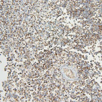
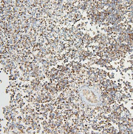
ARG45186 anti-VDAC3 antibody IHC-P image
Immunohistochemistry: Human testis cancer stained with ARG45186 anti-VDAC3 antibody at 2 μg/ml dilution.
-
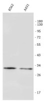
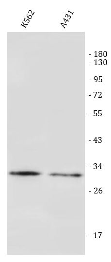
ARG45186 anti-VDAC3 antibody WB image
Western blot: K562 and A431 stained with ARG45186 anti-VDAC3 antibody at 0.5 μg/ml dilution.
-
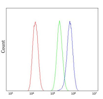
ARG45186 anti-VDAC3 antibody FACS image
Flow Cytometry: U20S stained with ARG45186 anti-VDAC3 antibody at 1 µg/10^6 cells dilution.
-
ARG45186 anti-VDAC3 antibody IHC-P image
Immunohistochemistry: Rat testis stained with ARG45186 anti-VDAC3 antibody at 2 μg/ml dilution.
-

ARG45186 anti-VDAC3 antibody WB image
Western blot: Rat heart and Rat kidney stained with ARG45186 anti-VDAC3 antibody at 0.5 μg/ml dilution.
-
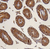
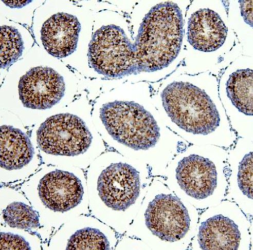
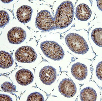
ARG45186 anti-VDAC3 antibody IHC-P image
Immunohistochemistry: Mouse testis stained with ARG45186 anti-VDAC3 antibody at 2 μg/ml dilution.
-
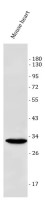
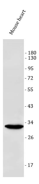
ARG45186 anti-VDAC3 antibody WB image
Western blot: Mouse heart stained with ARG45186 anti-VDAC3 antibody at 0.5 μg/ml dilution.