ARG30359
PINK1-Parkin Mitophagy Antibody Panel
Component
| Cat No | Component Name | Host clonality | Reactivity | Application | Package |
|---|---|---|---|---|---|
| ARG67305 | anti-PINK1 antibody | Rabbit pAb | Hu, Ms, Rat | WB | 20 μl |
| ARG58151 | anti-Parkin antibody | Rabbit pAb | Hu, Ms, Rat | FACS, ICC/IF, IHC-P, WB | 20 μl |
| ARG55799 | anti-LC3B antibody | Rabbit pAb | Hu, Ms, Rat | ICC/IF, IHC-P, WB | 20 μl |
| ARG66326 | anti-COX4 antibody [SQab1874] | Rabbit mAb | AGMK, Bov, Chk, Dog, Hu, Ms, Pig, Rat | FACS, ICC/IF, IHC-P, IP, WB | 20 μl |
Overview
| Product Description | PINK1-Parkin Mitophagy Antibody Panel is an all-in-one solution to make PINK1-Parkin Mitophagy research easy and economic. |
|---|---|
| Target Name | PINK1 / Parkin / LC3B / COX4 |
Properties
| Storage Instruction | -20°C |
|---|---|
| Note | For continuous use, store undiluted antibody at 2-8°C for up to a week. For long-term storage, aliquot and store at -20°C or below. Storage in frost free freezers is not recommended. Avoid repeated freeze/thaw cycles. Suggest spin the vial prior to opening. The antibody solution should be gently mixed before use. |
Bioinformation
| Gene Full Name | Antibody Panel for PINK1-Parkin Mitophagy |
|---|---|
| Highlight | Related Product: anti-PINK1 antibody; anti-Parkin antibody; anti-LC3B antibody; anti-COX4 antibody; |
Images (26) Click the Picture to Zoom In
-

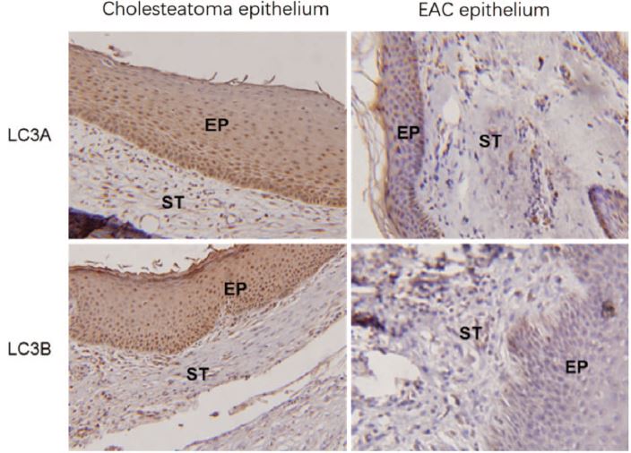
ARG55799 anti-LC3B antibody IHC-P image
Immunohistochemistry: Human epithelium stained with ARG51300 anti-LC3A antibody and ARG55799 anti-LC3B antibody.
From Li Q et al. Otol Neurotol (2019), doi: 10.1097/MAO.0000000000002404, Fig. 2.
-
ARG66326 anti-COX4 antibody [SQab1874] WB image
Western blot: THP-1 stained with ARG66326 anti-COX4 antibody [SQab1874].
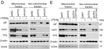
From Jian-Hong Shi et al. Cell Death Dis- (2022), doi: 10.1038/s41419-022-04539-9, Fig. 8. E.
-
ARG55799 anti-LC3B antibody ICC/IF image
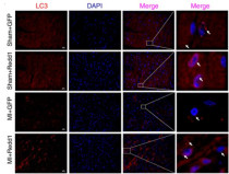
Immunofluorescence: Mouse heart stained with ARG55799 anti-LC3B antibody.
From Huang P et al. Int J Mol Med- (2019), doi: 10.3892/ijmm.2019.4366, Fig. 5. D.
-

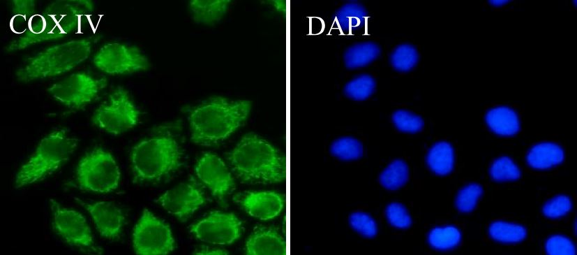
ARG66326 anti-COX4 antibody [SQab1874] ICC/IF image
Immunofluorescence: HeLa cells were fixed with 4% paraformaldehyde for 30 min at RT, permeabilized with 0.1% Triton X-100 for 10 min at RT then blocked with 10% goat serum for 30 min at RT. Cells were stained with ARG66326 anti-COX4 antibody [SQab1874] (green) at 1:50 and 4°C. DAPI (blue) was used as the nuclear counter stain.
-
ARG66326 anti-COX4 antibody [SQab1874] FACS image
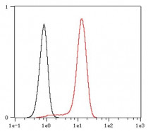
Flow Cytometry: HeLa cells were fixed with 4% paraformaldehyde (10 min) and then permeabilized with 0.1% TritonX-100 for 15 min. The cells were stained with ARG66326 anti-COX4 antibody [SQab1874] (red) at 1:1,000 dilution in 1x PBS/1% BSA for 30 min at RT, followed by Alexa Fluor® 488 labelled secondary antibody. Unlabelled sample (black) was used as a control.
-

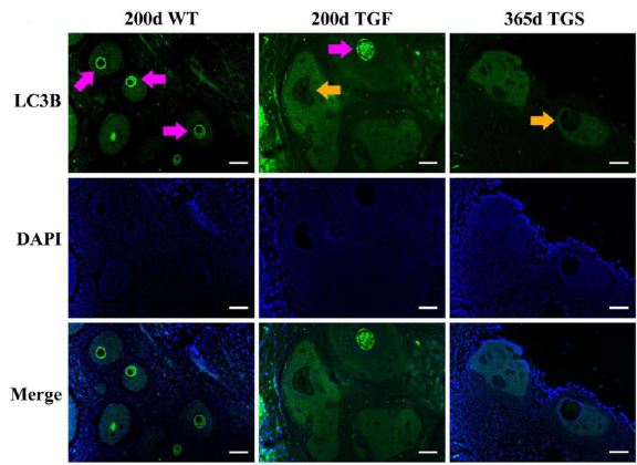
ARG55799 anti-LC3B antibody IHC-P image
Immunohistochemistry: Porcine ovarian stained with ARG55799 anti-LC3B antibody.
From Yufeng Qin et al. Preprint- (2019), doi: 10-1101-724096, Fig. 5. C.
-
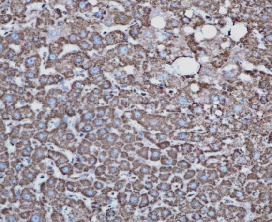
ARG66326 anti-COX4 antibody [SQab1874] IHC-P image
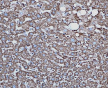
Immunohistochemistry: Formalin-fixed and paraffin-embedded liver tissue stained with ARG66326 anti-COX4 antibody [SQab1874] at 1:200 dilution. Antigen Retrieval: Heat mediated was performed using Tris/EDTA buffer pH 9.0.
-
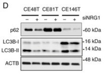
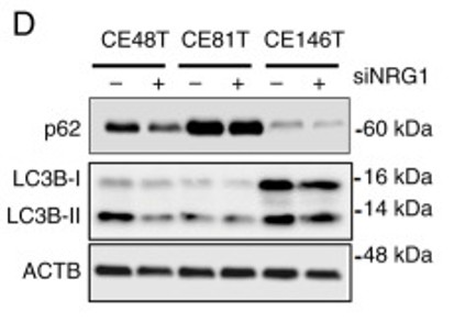
ARG55799 anti-LC3B antibody WB image
Western blot: ESCC cell stained with ARG55799 anti-LC3B antibody and ARG55040 anti-SQSTM1 / p62 antibody.
From Tseng YC et al. Int J Mol Med. (2025), doi: 10.3892/ijmm.2025.5503, Fig. 5. D.
-
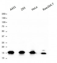
ARG66326 anti-COX4 antibody [SQab1874] WB image
Western blot: 10 µg of A431, 293, HeLa and Raw264.7 cell lysates stained with ARG66326 anti-COX4 antibody [SQab1874] at 1:5000 dilution.
-

ARG55799 anti-LC3B antibody WB image
Western blot: Mouse heart stained with ARG51300 anti-LC3A antibody and ARG55799 anti-LC3B antibody.
From Li Q et al. Otol Neurotol (2019), doi: 10.1097/MAO.0000000000002404, Fig. 2.
-
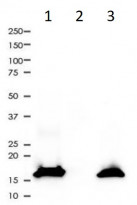
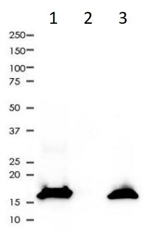
ARG66326 anti-COX4 antibody [SQab1874] IP image
Immunoprecipitation: 0.4 mg of HeLa whole cell lysate immunoprecipitated (1:50) and stained with ARG66326 anti-COX4 antibody [SQab1874]. 1) ARG66326 IP in HeLa whole cell lysate, 2) PBS instead of ARG66326 in HeLa whole cell lysate, and 3) HeLa whole cell lysate, 10 µg (input).
-
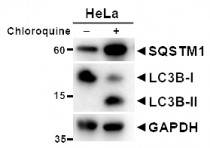
ARG55799 anti-LC3B antibody WB image (Customer's Feedback)
Western blot: HeLa cells untreated or treated with Chloroquine. 20 µg of cell lysates stained with ARG55040 anti-SQSTM1 / p62 antibody and ARG55799 anti-LC3B antibody at 1:1000 dilution.
An anti-GAPDH antibody used as a positive control.
-

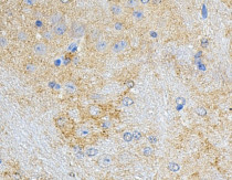
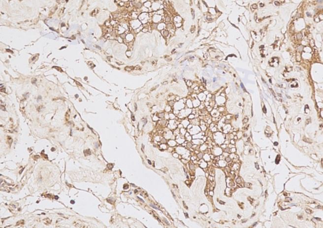
ARG58151 anti-Parkin antibody IHC-P image
Immunohistochemistry: Human testis stained with ARG58151 anti-Parkin antibody.
-

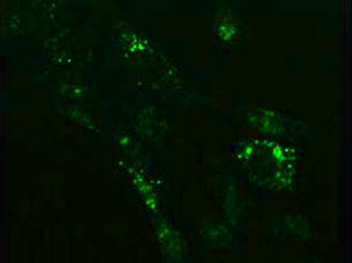
ARG55799 anti-LC3B antibody ICC/IF image
Immunofluorescence: NIH/3T3 cells treated with Chloroquine (50 μM, 37°C for 20 hours). Cells were stained with ARG55799 anti-LC3B antibody at 1:100 dilution.
-

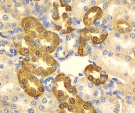
ARG55799 anti-LC3B antibody IHC-P image
Immunohistochemistry: Paraffin-embedded Rat kidney stained with ARG55799 anti-LC3B antibody at 1:100 dilution.
-
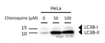
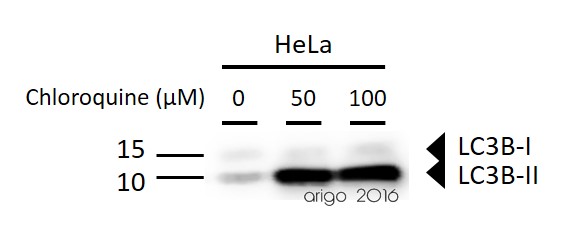
ARG55799 anti-LC3B antibody WB image
Western blot: 30 µg of HeLa untreated or treated with Chloroquine at 50 µM, 100 µM (18 hr). The bolts stained with ARG55799 anti-LC3B antibody at 1:1000 dilution.
-
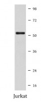
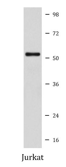
ARG58151 anti-Parkin antibody WB image
Western blot: Jurkat cell lysate stained with ARG58151 anti-Parkin antibody.
-
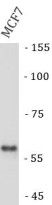
ARG67305 anti-PINK1 antibody WB image
Western blot: MCF7 stained with ARG67305 anti-PINK1 antibody.
-
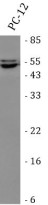
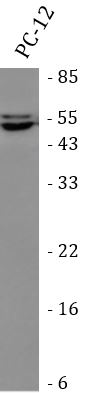
ARG58151 anti-Parkin antibody WB image
Western blot: PC-12 stained with ARG58151 anti-Parkin antibody.
-
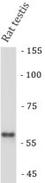
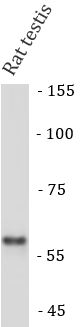
ARG67305 anti-PINK1 antibody WB image
Western blot: Rat testis stained with ARG67305 anti-PINK1 antibody.
-
ARG58151 anti-Parkin antibody IHC-P image
Immunohistochemistry: Mouse brain stained with ARG58151 anti-Parkin antibody.
-
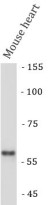
ARG58151 anti-Parkin antibody WB image
Western blot: Mouse brain stained with ARG58151 anti-Parkin antibody.
-
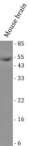
ARG67305 anti-PINK1 antibody WB image
Western blot: Mouse heart stained with ARG67305 anti-PINK1 antibody.
-

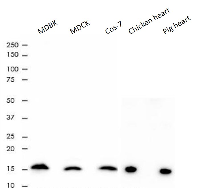
ARG66326 anti-COX4 antibody [SQab1874] WB image
Western blot: 10 µg of MDBK, MDCK, Cos-7, Chicken heart and Pig heart lysates stained with ARG66326 anti-COX4 antibody [SQab1874] at 1:5000 dilution.
-

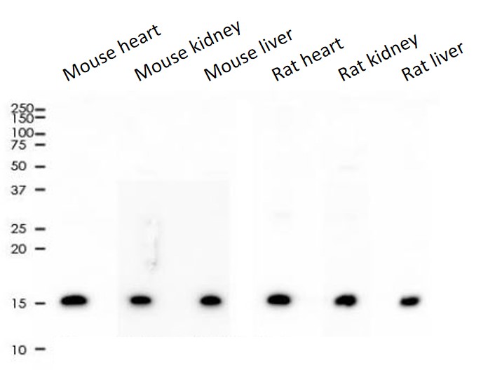
ARG66326 anti-COX4 antibody [SQab1874] WB image
Western blot: 2 µg of Mouse heart, Mouse kidney, Mouse liver, Rat heart, Rat kidney and Rat liver lysates stained with ARG66326 anti-COX4 antibody [SQab1874] at 1:5000 dilution.
-
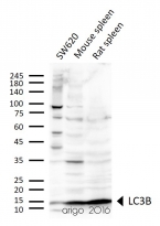
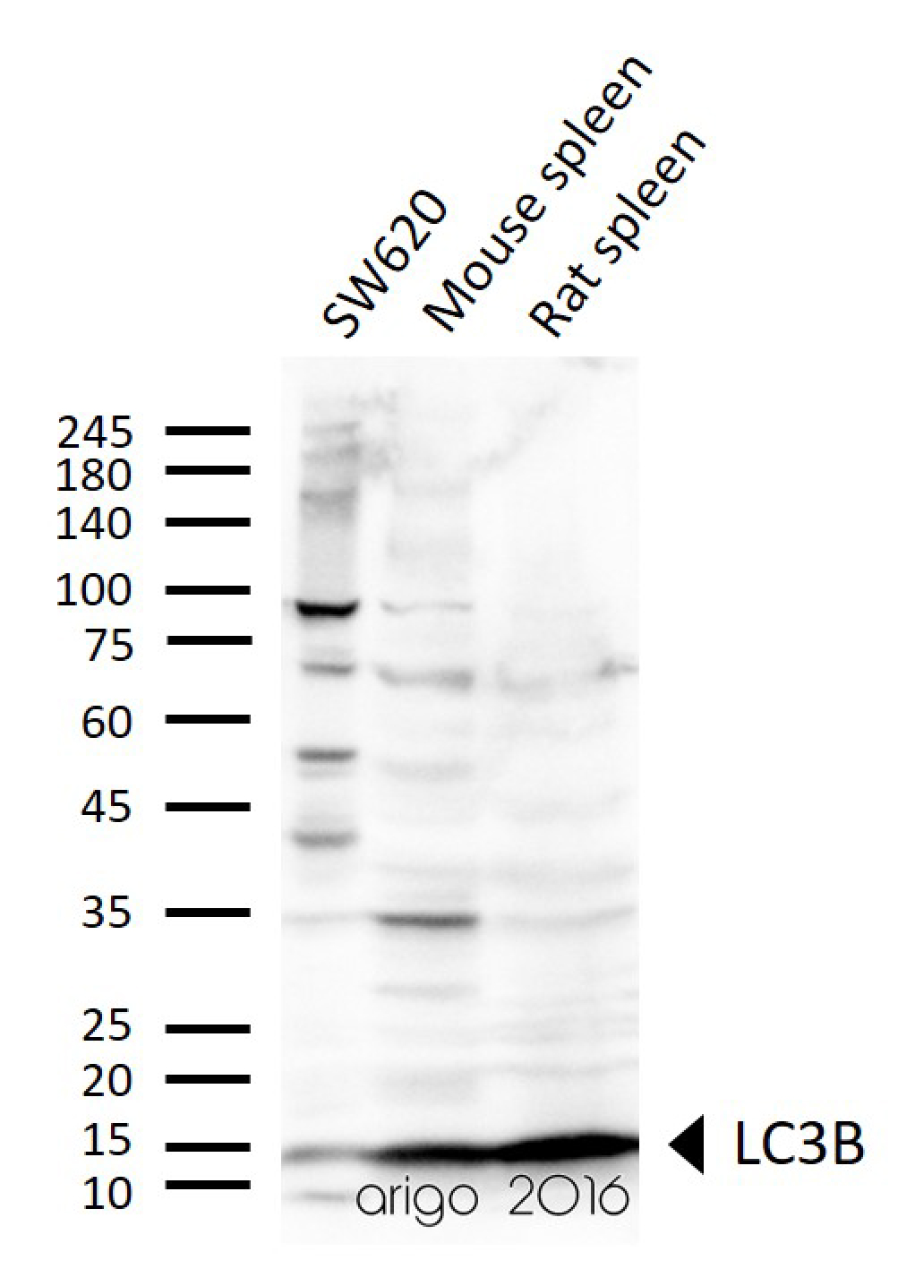
ARG55799 anti-LC3B antibody WB image
Western blot: 30 µg of SW620, Mouse spleen, and Rat spleen lysates stained with ARG55799 anti-LC3B antibody at 1:1000 dilution.


Specific References
Ailanthone targets the KMT2A-MEN1 complex to suppress lung metastasis of osteosarcoma
ARG55799; WB / Human


