Detecting exosomal PD-L1 secreted by cancer cells
Detecting exosomal PD-L1 secreted by cancer cells
Exosomes is the extracellular vesicles secreted by all cells. Evidences accumulated that exosomes implicate in various diseases including cancer metastasis and neurodegeneration diseases. PD-L1 is an immune checkpoint inhibitor that keep T cells from attacking other cells in the body. Many cancer cells overexpress PD-L1 to escape the immunological attack.
The exosomal PD-L1 has been isolated in multiple cancer cells. Recently, Chen et al. elucidated the regulation and immunosuppressive function of exosomal PD-L1 and published their result in Nature. They found that IFNγ treatment enhances the levels of PD-L1 in exosomes. In mouse model, exosomal PD-L1 inhibits CD8+ T cells and facilitates the cancer progression. Moreover, the authors examined the exosomel PD-L1 levels in melanoma patients during anti-PD-1 therapy and found that the pretreatment level of exosomal PD-L1 was higher in non-responders which was associated with poor prognosis. Their study provides a rationale for the application of exosomal PD-L1 as a predictor for anti-PD-1 therapy.
arigo offers excellent ELISA kits to detect exosomal PD-L1 level. In addition, more quality antibodies and ELISA kits are available for facilitating related studies.
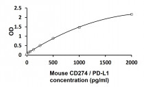
● Detecting exosomal PD-L1
| Human PD-L1 ELISA Kit (ARG81929) Sensitivity: 31.25 pg/ml Samples: Serum, Plasma, Cell culture sup. 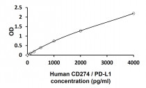 |
Mouse PD-L1 ELISA Kit (ARG81930) |
● Exosome Antibody Panel (ARG30348)
|
- Includes exosome marker CD63 antibody for detecting tetraspanin |
| Component | Reactivity | Application | Pkg |
| CD63 antibody | Hu | ICC/IF, IHC-P, WB | 20 ul |
| TSG101 antibody | Hu, Ms, Rat | FACS, ICC/IF, IHC-P, WB | 20 ul |
| GM130 antibody | Hu, Ms, Rat, Cow, Dog, Mk | ICC/IF, IHC-P, IP, WB | 20 ul |
Reference: Chen et al., (2018) Nature. 560(7718):382-386.
