ARG30358
Mitochondrial Biogenesis Antibody Panel
Component
| Cat No | Component Name | Host clonality | Reactivity | Application | Package |
|---|---|---|---|---|---|
| ARG59247 | anti-PGC1 alpha antibody | Rabbit pAb | Hu, Ms, Rat | ICC/IF, WB | 20 μl |
| ARG58207 | anti-NRF1 antibody | Rabbit pAb | Hu, Ms, Rat | FACS, ICC/IF, IHC-P, IP, WB | 20 μl |
| ARG46741 | anti-TFAM antibody | Rabbit pAb | Hu, Ms, Rat | ICC/IF, IHC-P, WB | 20 μl |
Overview
| Product Description | Mitochondrial Biogenesis Antibody Panel is an all-in-one solution to make Mitochondrial Biogenesis research easy and economic. |
|---|---|
| Target Name | PGC1 alpha / NRF1 / TFAM |
Properties
| Storage Instruction | -20°C |
|---|---|
| Note | For continuous use, store undiluted antibody at 2-8°C for up to a week. For long-term storage, aliquot and store at -20°C or below. Storage in frost free freezers is not recommended. Avoid repeated freeze/thaw cycles. Suggest spin the vial prior to opening. The antibody solution should be gently mixed before use. |
Bioinformation
| Gene Full Name | Antibody Panel for Mitochondrial Biogenesis |
|---|---|
| Highlight | Related Product: anti-PGC1 alpha antibody; anti-NRF1 antibody; anti-TFAM antibody; |
Images (15) Click the Picture to Zoom In
-

ARG58207 anti-NRF1 antibody WB image
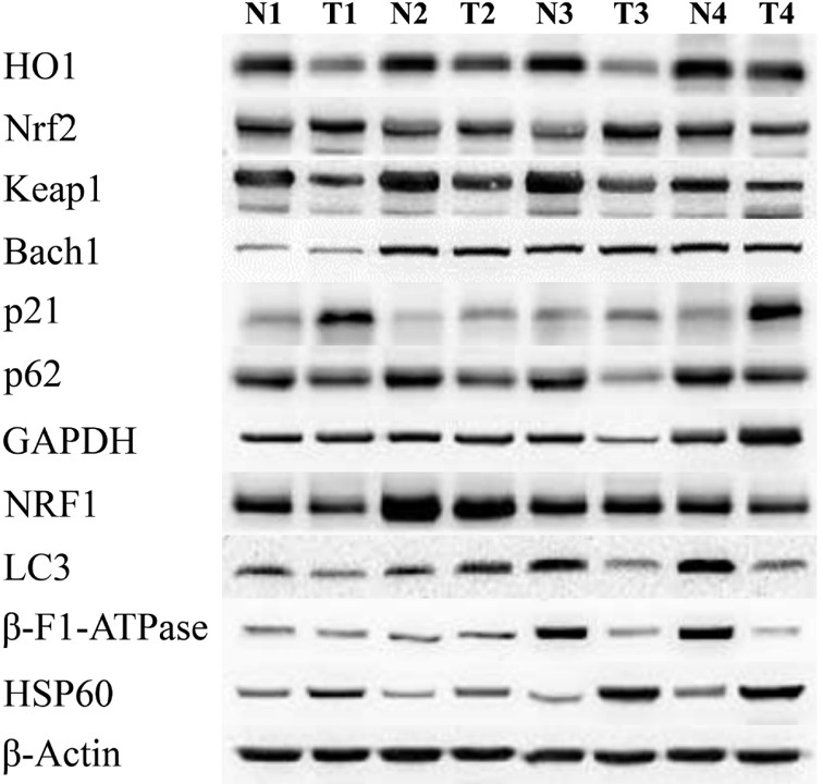
Western blot: Human CRC stained with ARG58207 anti-NRF1 antibody.
From Chang LC et al. Cancer Biomark- (2020), doi: 10.3233/CBM-190828, Fig. 2.
-

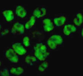
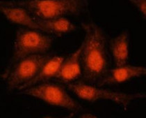
ARG58207 anti-NRF1 antibody ICC/IF image
Immunofluorescence: 293T cells stained with ARG58207 anti-NRF1 antibody at 1:250 dilution.
-

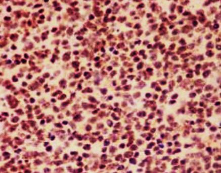
ARG58207 anti-NRF1 antibody IHC-P image
Immunohistochemistry: Paraffin-embedded tonsil tissue stained with ARG58207 anti-NRF1 antibody.
-
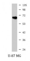
ARG58207 anti-NRF1 antibody WB image
Western blot: U-87 MG cell lysate stained with ARG58207 anti-NRF1 antibody.
-
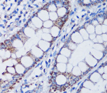
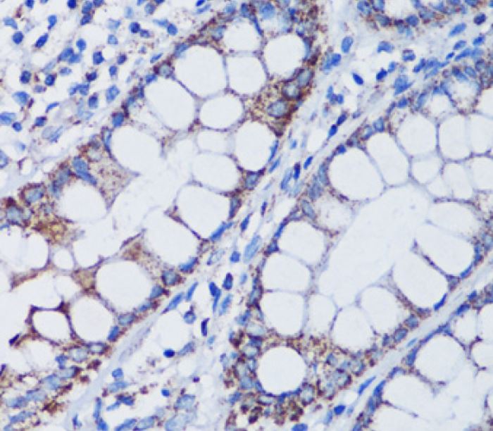
ARG46741 anti-TFAM antibody IHC-P image
Immunohistochemistry: Human colon stained with ARG46741 anti-TFAM antibody.
-
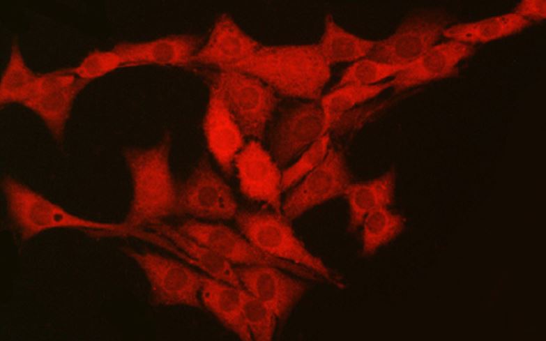
ARG59247 anti-PGC1 alpha antibody ICC/IF image
Immunofluorescence: HeLa cells stained with ARG59247 anti-PGC1 alpha antibody at 1:100 dilution.
-
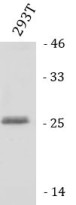
ARG59247 anti-PGC1 alpha antibody WB image
Western blot: 25 µg of 293T cell lysate stained with ARG59247 anti-PGC1 alpha antibody at 1:1000 dilution.
-
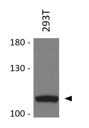
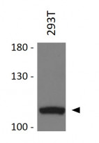
ARG46741 anti-TFAM antibody WB image
Western blot: 293T stained with ARG46741 anti-TFAM antibody.
-
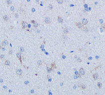
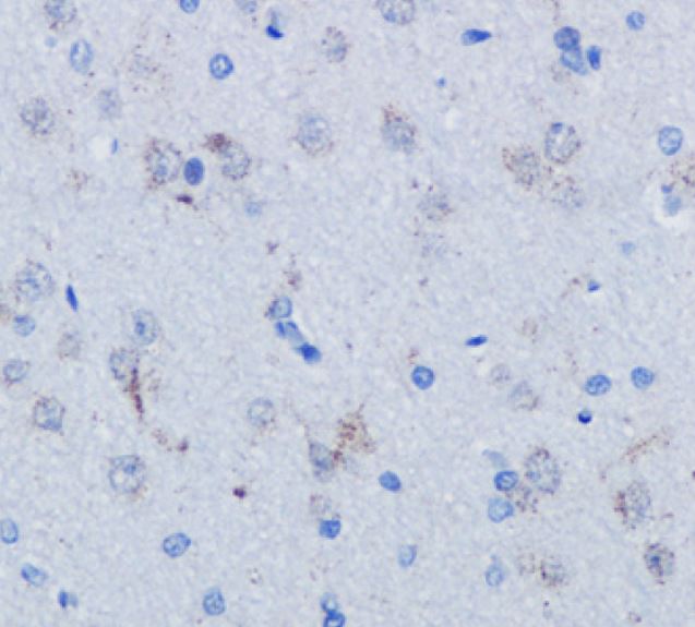
ARG46741 anti-TFAM antibody IHC-P image
Immunohistochemistry: Rat brain stained with ARG46741 anti-TFAM antibody.
-

ARG59247 anti-PGC1 alpha antibody IHC-P image
Immunohistochemistry: PC-12 stained with ARG59247 anti-PGC1 alpha antibody.
-
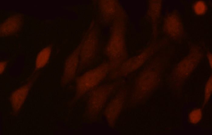
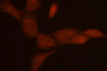
ARG46741 anti-TFAM antibody ICC/IF image
Immunofluorescence: PC-12 stained with ARG46741 anti-TFAM antibody at dilution.
-
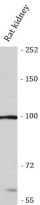
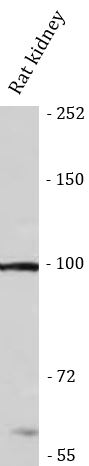
ARG59247 anti-PGC1 alpha antibody WB image
Western blot: Rat kidney stained with ARG59247 anti-PGC1 alpha antibody.
-
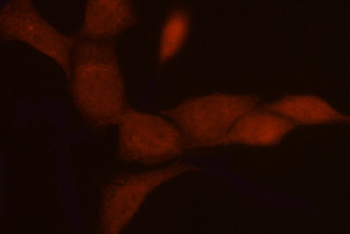
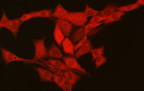
ARG59247 anti-PGC1 alpha antibody ICC/IF image
Immunofluorescence: NIH/3T3 stained with ARG59247 anti-PGC1 alpha antibody at dilution.
-

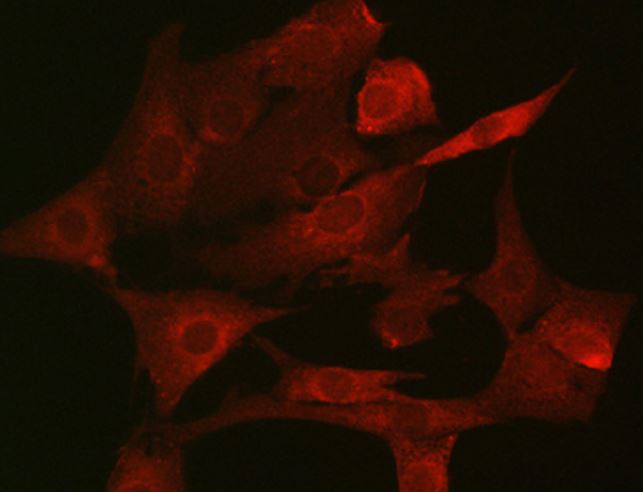
ARG46741 anti-TFAM antibody ICC/IF image
Immunofluorescence: NIH/3T3 stained with ARG46741 anti-TFAM antibody at dilution.
-
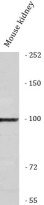
ARG59247 anti-PGC1 alpha antibody WB image
Western blot: Mouse kidney stained with ARG59247 anti-PGC1 alpha antibody.