ARG30170
Phospho Src Antibody Panel
Cancer antibody; Gene Regulation antibody; Immune System antibody; Metabolism antibody; Signaling Transduction antibody
Component
| Cat No | Component Name | Host clonality | Reactivity | Application | Package |
|---|---|---|---|---|---|
| ARG54712 | anti-Src antibody [17AT28] | Mouse mAb | Hu, Ms | ICC/IF, WB | 50 μl |
| ARG51594 | anti-Src phospho (Tyr418) antibody | Rabbit pAb | Hu, Ms, Rat | IHC-P, WB | 50 μl |
| ARG51652 | anti-Src phospho (Tyr529) antibody | Rabbit pAb | Hu, Ms, Rat | ICC/IF, IHC-P, WB | 50 μl |
| ARG65350 | Goat anti-Mouse IgG antibody (HRP) | Goat pAb | Ms | ELISA, IHC-P, WB | 50 μl |
| ARG65351 | Goat anti-Rabbit IgG antibody (HRP) | Goat pAb | Rb | ELISA, IHC-P, WB | 50 μl |
Overview
| Product Description | Src and Src-family protein-tyrosine kinases are regulatory proteins that play key roles in cell differentiation, motility, proliferation, and survival. The initially described phosphorylation sites of Src include an activating phosphotyrosine 418 that results from autophosphorylation, and an inhibiting phosphotyrosine 529 that results from phosphorylation by C-terminal Src kinase (Csk) and Csk homologous kinase. Dephosphorylation of phosphotyrosine 529 increases Src kinase activity. This antibody panel can be used for determine the activity of Src by different specific phosphorylation site. Wu S.S. et al. (2005) Cellular Signalling 17, 93-102. Obergfell A. et al. (2002) JCB 157, 265-275. Zhao M. et al. (2006) Mol. Cell. Biol. 26, 2479-2489. |
|---|---|
| Target Name | Src |
| Alternate Names | Phospho Src antibody; Src phospho (Tyr418) antibody; Src phospho (Tyr529) antibody; Src antibody |
Properties
| Storage Instruction | For continuous use, store undiluted antibody at 2-8°C for up to a week. For long-term storage, aliquot and store at -20°C or below. Storage in frost free freezers is not recommended. Avoid repeated freeze/thaw cycles. Suggest spin the vial prior to opening. The antibody solution should be gently mixed before use. |
|---|---|
| Note | For laboratory research only, not for drug, diagnostic or other use. |
Bioinformation
| Gene Full Name | Antibody Panel for Phospho Src |
|---|---|
| Highlight | Related Product: anti-Src antibody; |
| Research Area | Cancer antibody; Gene Regulation antibody; Immune System antibody; Metabolism antibody; Signaling Transduction antibody |
Images (17) Click the Picture to Zoom In
-
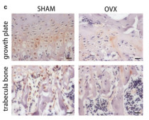
ARG65351 Goat anti-Rabbit IgG antibody (HRP) IHC-P image
From Yu-Qian Song et al. J Mol Med (Berl) (2022), doi: 10.1007/s00109-021-02165-0, Fig. 5.c.
-

ARG65351 Goat anti-Rabbit IgG antibody (HRP) WB image
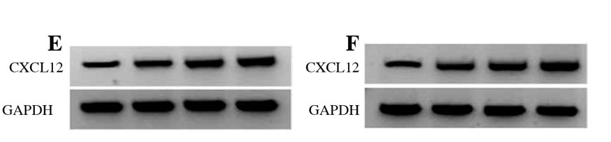
Western blot: HK2 stained with ARG66421 anti-CXCL12 / SDF1 antibody [SQab18123] and ARG10112 anti-GAPDH antibody [6C5] and secondary antibody used ARG65351 Goat anti-Rabbit IgG antibody (HRP).
From Zhou Y et al. Central European Journal of Immunology (2022), doi: 10.5114/ceji.2022.115628, Fig. 2. E.
-
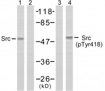
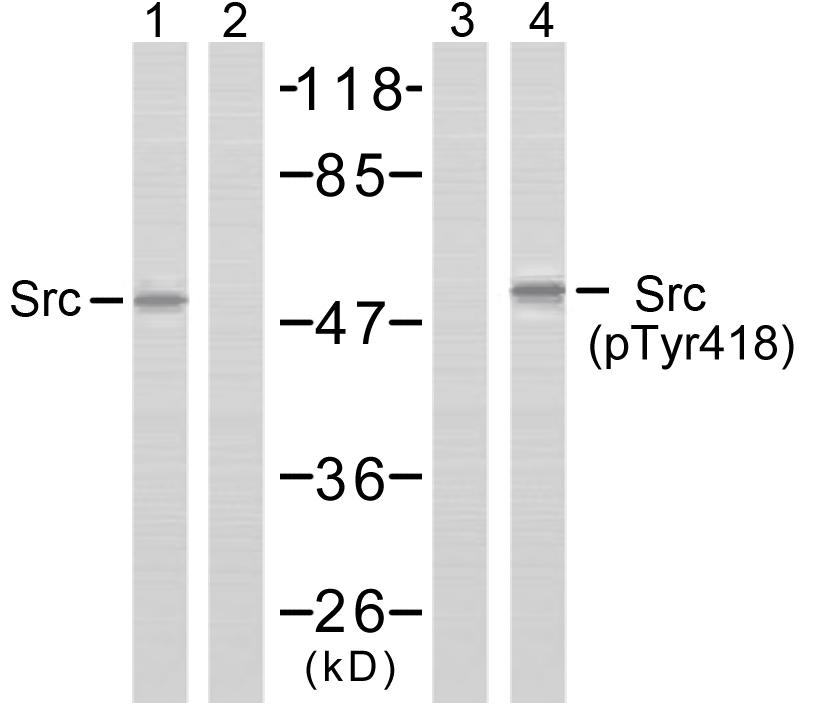
ARG51594 anti-Src phospho (Tyr418) antibody WB image
Western Blot: extracts from COLO205 cells stained with Src antibody (Lane 1 and 2) and Src (phospho-Tyr418) antibody (ARG51594, Lane 3 and 4).
-
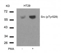
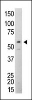
ARG51652 anti-Src phospho (Tyr529) antibody WB image
Western Blot: extracts from HT29 cells untreated or treated with PMA stained with anti-Src (phospho Tyr529) antibody ARG51652.
-
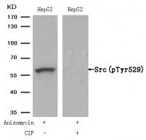
ARG51652 anti-Src phospho (Tyr529) antibody WB image
Western Blot: extracts from HepG2 cells, treated with Anisomycin or calf intestinal phosphatase (CIP), stained with anti-Src (phospho Tyr529) antibody ARG51652.
-
ARG54712 anti-SRC Antibody WB image
Western blot: Jurkat cell lysate stained with ARG54712 anti-SRC antibody.
-
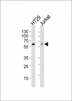
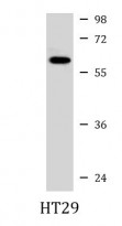
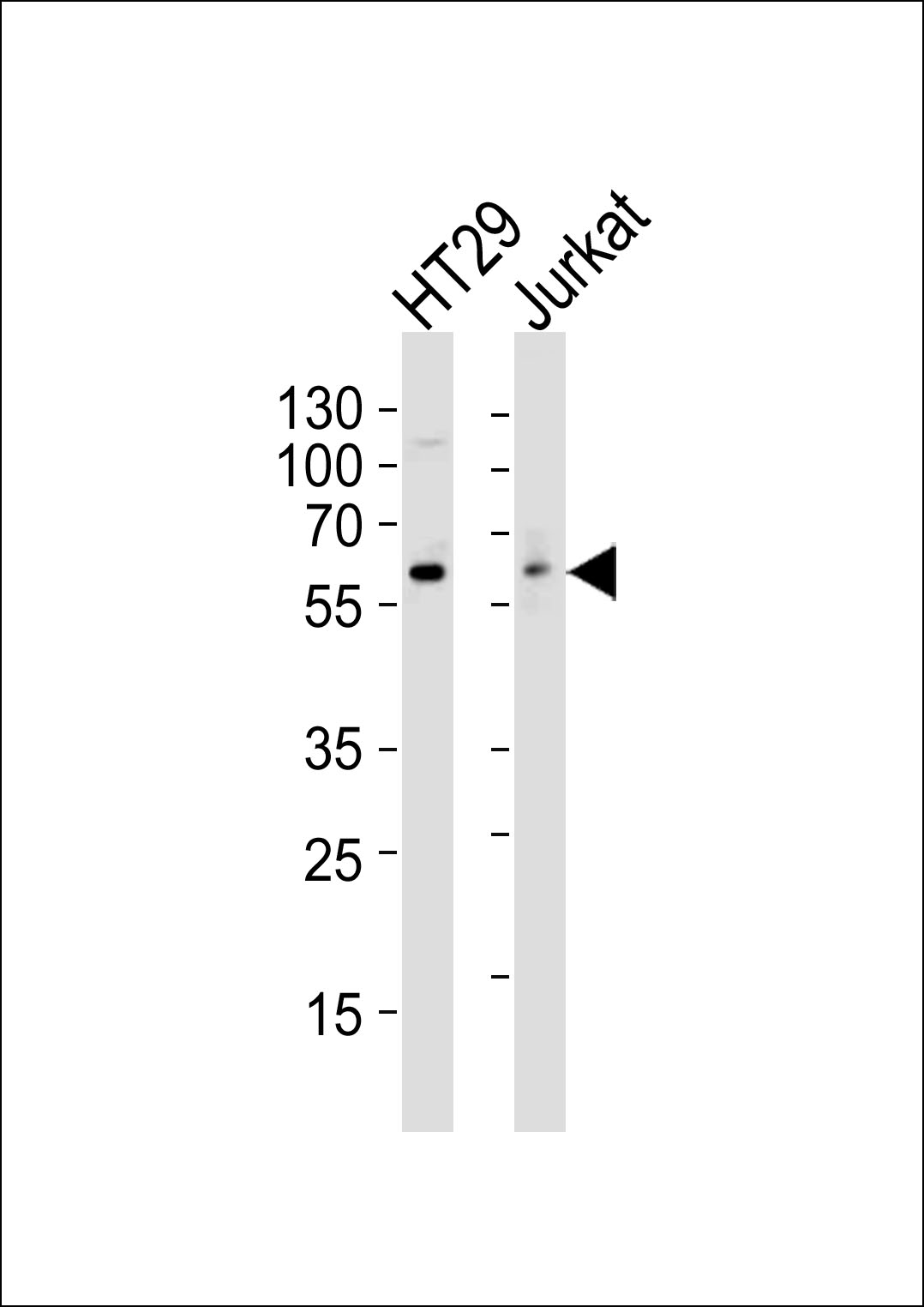
ARG54712 anti-SRC Antibody WB image
Western blot: 35 μg of HT29, Jurkat cell line lysates (from left to right) stained with ARG54712 anti-SRC antibody at 1:1000 dilution.
-

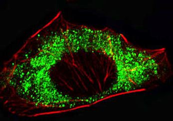
ARG54712 anti-SRC Antibody ICC/IF image
Immunofluorescence: A549 cells were fixed with 4% PFA (20 min), permeabilized with Triton X-100 (0.1%, 10 min), then stained with ARG54712 anti-SRC antibody (1:25, 1 h at 37°C). Cytoplasmic actin was counterstained with Alexa Fluor® 555 (red) conjugated Phalloidin (7 units/ml, 1 h at 37°C).
-
ARG51594 anti-Src phospho (Tyr418) antibody IHC-P image
Immunohistochemistry: paraffin- embedded human breast carcinoma tissue stained with anti-Src (phospho Tyr418) antibody ARG51594).
-
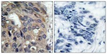
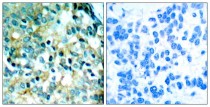
ARG51652 anti-Src phospho (Tyr529) antibody IHC-P image
Immunohistochemistry: paraffin-embedded human breast carcinoma tissue stained with anti-Src (phospho Tyr529) antibody ARG51652 (left) or the same antibody preincubated with blocking peptide (right).
-
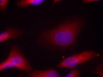
ARG51652 anti-Src phospho (Tyr529) antibody ICC/IF image
Immunofluorescence: methanol-fixed HeLa cells stained with anti-Src (phospho Tyr529) antibody ARG51652.
-
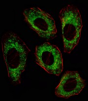
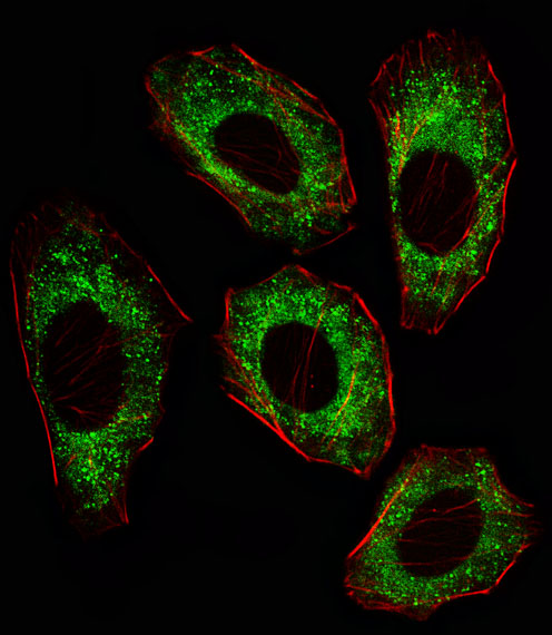
ARG54712 anti-SRC Antibody IF image
Immunofluorescence: A549 cell stained with ARG54712 anti-SRC antibody. A549 cells were fixed with 4% PFA (20 min), permeabilized with Triton X-100 (0.1%, 10 min), then incubated with SRC primary antibody (1:25, 1 h at 37°C). Cytoplasmic actin was counterstained with Alexa Fluor® 555 (red) conjugated Phalloidin (7units/ml, 1 h at 37°C). SRC immunoreactivity is localized to Cytoplasm significantly.
-
ARG54712 anti-SRC Antibody WB image
Western blot: 35 µg of HT29 cell lysate stained with ARG54712 anti-SRC antibody at 1:1000 dilution.
-
ARG65350 Goat anti-Mouse IgG antibody (HRP) IHC-P image
From Cheng-Feng Chu et al. J Pers Med. (2021), doi: 10.3390/jpm11121326, Fig. 6.
-
ARG65350 Goat anti-Mouse IgG antibody (HRP) WB image
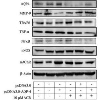
Western blot: Neuro-2a stained with ARG22361 anti-Aquaporin 4 antibody , ARG40638 anti-MMP9 antibody and ARG65350 Goat anti-Mouse IgG antibody (HRP).
From Hung CY et al. Molecules (2022), doi: 10.3390/molecules27031066, Fig. 6A.
-
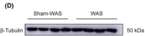
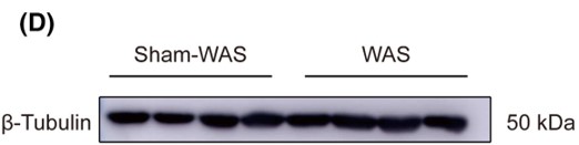
ARG65350 Goat anti-Mouse IgG antibody (HRP) WB image
Western blot: Rat basolateral amygdala stained with ARG62347 anti-beta Tubulin antibody [BT7R] at 1:1000 dilution, ARG65350 Goat anti-Mouse IgG antibody (HRP) at 1:5000 dilution.
From Guang-Bing Duan et al. CNS Neurosci Ther. (2024), doi: 10.1111/cns.14611, Fig. 4.D.
-
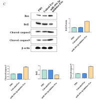
ARG65351 Goat anti-Rabbit IgG antibody (HRP) WB image
Western blot: Gastric cancer cells stained with ARG66247 anti-Bax antibody, ARG55188 anti-Bcl 2 antibody, ARG57512 anti-Caspase 3 (cleaved) antibody and ARG62346 anti-beta Actin antibody [BA3R].
Secondary Antibody stained with ARG65351 Goat anti-Rabbit IgG antibody (HRP).From Limin Zhang et al. Heliyon (2024), doi: 10.1016/j.heliyon.2024.e30803, Fig. 4. C.


Specific References
Bioactive Compounds of Momordica charantia L. Downregulate the Protein Expression of ACE2 and TMPRSS2 In Vivo and In Vitro
ARG65351; WB /


γ-Terpinene from citrus peel enhances mitochondrial bioenergetics and protects neuronal cells from glutamate-induced injury
ARG65350; WB / Rat
Study on the Role of Short Peptide LCKLSL Targeting ANXA2 in Retinal Neovascularization
ARG65350; WB /
Protective efficacy of the recombinantly expressed C-terminal domain of Mannheimia haemolytica leukotoxin in mice and goats
ARG65350: WB /
Mechanical and interfacial properties of zwitterionic hydrogels via a high-entanglement network design: a study of monomer and crosslinker synergy
ARG65351: FuncSt /
Exosomes separation with aqueous two-phase systems from bovine milk
ARG65350; WB /
A New Way to Engineer Cell Sheets forArticular Cartilage Regeneration
ARG65350; ICC/IF /
Soybean Trypsin Inhibitor Possesses Potency Against SARS-CoV-2 Infection by Blocking the Host Cell Surface Receptors ACE2, TMPRSS2, and CD147
ARG65351; WB /
Reduced endometrial glycolysis concomitant with increased lesional fibrosis in patients with adenomyosis who complained of heavy menstrual bleeding
ARG65351; WB /
Progressively diminished estrogen signaling concordant with increased fibrosis in ectopic endometrium
ARG65351; WB /
Calycosin Induces Ferroptosis by SLC7A11 Through the PI3K/Akt Pathway in Acute Myelocytic Leukemia
ARG65351; WB /
Chronic intranasal oxytocin alleviates cognitive impairment and reverses oxytocin signaling upregulation in MK801-induced mice
ARG65351; WB /
Overexpression of ZNF468 promotes esophageal squamous cell carcinoma progression via the AKT/mTOR pathway
ARG65351; WB /
Anti-CTLA-4 antibody self-presented dendritic cell nanovesicles boost the immunotherapy of hepatocellular carcinoma after microwave ablation
ARG65350; WB /
Expression and correlation of Surfeit 4 gene in esophageal squamous cell carcinoma
ARG65350; WB /
Blood RNA-sequencing analysis in acrylamide-induced neurotoxicity and depressive symptoms in rats
ARG65350; WB /
The Therapeutic Potential of Exosomes vs. Matrix-Bound Nanovesicles from Human Umbilical Cord Mesenchymal Stromal Cells in Osteoarthritis Treatment
ARG65351; WB /
Four amino acids play an important role in the allergenicity of hemocyanin allergen
ARG65350; WB /
Environmental acidification drives inter-organ energy mobilization to enhance reproductive performance in medaka (Oryzias latipes)
ARG65351; WB /
KDF1 Promoted Proliferation, Migration and Invasion of Lung Adenocarcinoma Cells through Activating STAT3 and AKT Pathway
ARG65350: WB /


