ARG30330
Pyroptosis Antibody Panel
Component
| Cat No | Component Name | Host clonality | Reactivity | Application | Package |
|---|---|---|---|---|---|
| ARG57293 | anti-Caspase 1 antibody | Rabbit pAb | Hu, Ms | ICC/IF, IHC-P, IP, WB | 20 μl |
| ARG41404 | anti-GSDMD antibody | Rabbit pAb | Hu, Ms, Rat | ICC/IF, WB | 20 μl |
| ARG66285 | anti-IL1 beta antibody [SQab1748] | Mouse mAb | Hu, Ms | ELISA, FACS, ICC/IF, IHC-P, WB | 20 μg |
| ARG62347 | anti-beta Tubulin antibody [BT7R] | Mouse mAb | Hu, Ms, Rat, Chk, Mk, Rb | Dot, ELISA, ICC/IF, WB | 20 μg |
Overview
| Product Description | Pyroptosis Antibody Panel is an all-in-one solution to make pyroptosis research easy and economic. This antibody panel comprises antibodies against pyroptosis key players Caspase-1, GSDMD, and IL1 beta. All three antibodies are good for detecting both pro- and cleaved proteins. Moreover, a loading control antibody against Tubulin is also included. This panel is the best solution for studying pyroptosis. Related news: Inflammasome & Pyroptosis Antibody Panels are released; Exploring Antiviral Immune Response; RIP1 activation and pathogenesis of NASH; AMPK signaling regulates NLRP3 inflammation and pyroptosis; Solutions for studying PANoptosis & PANoptosome; m6A reader YTHDF2 in mRNA decay and aggresome formation; |
|---|---|
| Target Name | Pyroptosis |
| Alternate Names | Pyroptosis antibody; GSDMD antibody; Caspase 1 antibody; beta Tubulin antibody; IL1 beta antibody |
Properties
| Storage Instruction | For continuous use, store undiluted antibody at 2-8°C for up to a week. For long-term storage, aliquot and store at -20°C or below. Storage in frost free freezers is not recommended. Avoid repeated freeze/thaw cycles. Suggest spin the vial prior to opening. The antibody solution should be gently mixed before use. |
|---|---|
| Note | For laboratory research only, not for drug, diagnostic or other use. |
Bioinformation
| Gene Full Name | Antibody Panel for Pyroptosis |
|---|---|
| Highlight | Related Product: anti-Caspase 1 antibody; anti-GSDMD antibodies; anti-IL1 beta antibody; |
Images (26) Click the Picture to Zoom In
-
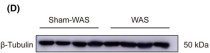
ARG62347 anti-beta Tubulin antibody [BT7R] WB image
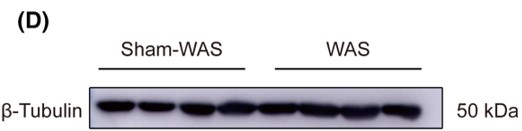
Western blot: Rat basolateral amygdala stained with ARG62347 anti-beta Tubulin antibody [BT7R] at 1:1000 dilution, ARG65350 Goat anti-Mouse IgG antibody (HRP) at 1:5000 dilution.
From Guang-Bing Duan et al. CNS Neurosci Ther. (2024), doi: 10.1111/cns.14611, Fig. 4.D.
-

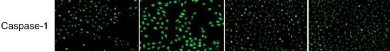
ARG57293 anti-Caspase 1 antibody ICC/IF image
Immunofluorescence: HUVECs stained with ARG57293 anti-Caspase 1 antibody.
From Yuan Y et al. Mol Med Rep (2022), doi: 10.3892/mmr.2022.12730, Fig. 3B.
-

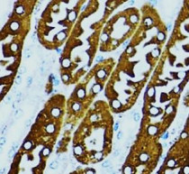
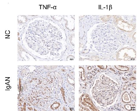
ARG66285 anti-IL1 beta antibody [SQab1748] IHC-P image
Immunohistochemistry: Human renal stained with ARG10158 anti-TNF alpha antibody [2C8] and ARG66285 anti-IL1 beta antibody [SQab1748].
From Tan YY et al. Ren Fail (2025), doi: 10.1080/0886022X.2025.2508297, Fig. 6A.
-
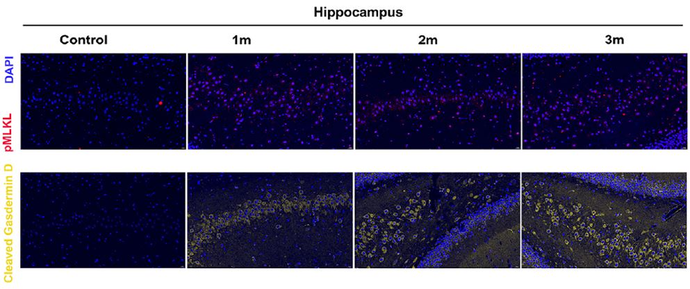
ARG41404 anti-GSDMD antibody IHC-P image
Immunohistochemistry: Rat hippocampus stained with ARG41404 anti-GSDMD antibody and ARG43708 anti-MLKL antibody at 1:100 dilution.
From Wu M et al. Sci Rep (2025), doi: 10.1038/s41598-025-01087-y, Fig. 2B.
-
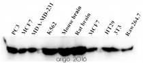
ARG62347 anti-beta Tubulin antibody [BT7R] WB image
Western blot: 20 μg of PC3, MCF7, MDA-MD-231, K562, M. brain, R. brain, MCF7, HT29, 3T3 and Raw264.7 cell lysates stained with ARG62347 anti-beta Tubulin antibody [BT7R] at 1:3000 dilution.
-
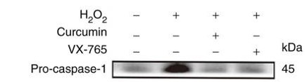
ARG57293 anti-Caspase 1 antibody WB image
Western blot: HUVECs stained with ARG57293 anti-Caspase 1 antibody at 1:1000 dilution.
From Yulin Yuan et al. Mol Med Rep. (2022), doi: 10.3892/mmr.2022.12730, Fig. 3.(A).
-
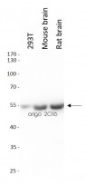
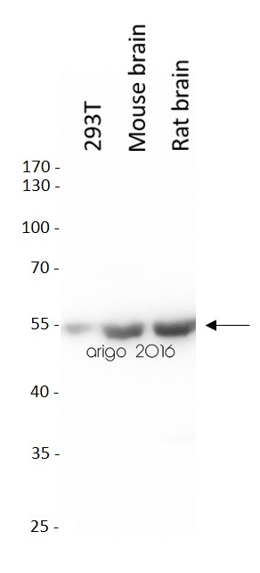
ARG62347 anti-beta Tubulin antibody [BT7R] WB image
Western blot: 20 µg of 293T, Mouse brain and Rat brain lysates stained with ARG62347 anti-beta Tubulin antibody [BT7R] at 1:10000 dilution.
-

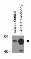
ARG57293 anti-Caspase 1 antibody WB image
Western blot: Mouse liver stained with ARG57293 anti-Caspase 1 antibody.
From Zhao Y et al. Preprint- (2021), doi: 10.1038/s41598-021-00071-6, Fig. 4F.
-

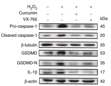
ARG41404 anti-GSDMD antibody WB image
Western blot: HUVECs stained with ARG41404 anti-GSDMD antibody , and ARG66285 anti-IL1 beta antibody [SQab1748]at 1:100 dilution.
From Yulin Yuan et al. Mol Med Rep. (2022), doi: 10.3892/mmr.2022.12730, Fig. 3.(A).
-
ARG41404 anti-GSDMD antibody WB image
Western blot: Rat brain stained with ARG41404 anti-GSDMD antibody and ARG10644 anti-Caspase 1 antibody .
From Wu M et al. Sci Rep (2025), doi: 10.1038/s41598-025-01087-y, Fig. 3A.
-

ARG41404 anti-GSDMD antibody WB image
Western blot: Mouse liver stained with ARG41404 anti-GSDMD antibody.
From Meng Z et al. J Agric Food Chem (2023), doi: 10.1021/acs.jafc.2c07581, Fig. 2B.
-
ARG41404 anti-GSDMD antibody WB image
Western blot: Mouse liver stained with ARG41404 anti-GSDMD antibody and ARG66285 anti-IL1 beta antibody [SQab1748] .
From Lu J et al. Food Chem Toxicol. (2023), doi: 10.1016/j.fct.2023.114060, Fig. 1. D.
-
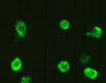
ARG41404 anti-GSDMD antibody ICC/IF image
Immunofluorescence: RAW264.7 cells stained with ARG41404 anti-GSDMD antibody at 1:100 dilution.
-
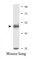
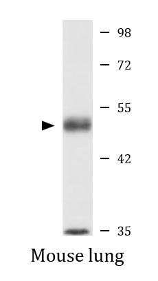
ARG41404 anti-GSDMD antibody WB image
Western blot: 25 µg of Mouse lung lysate stained with ARG41404 anti-GSDMD antibody at 1:1000 dilution.
-

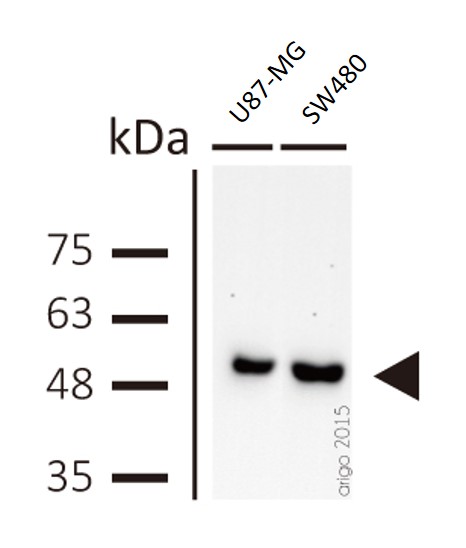
ARG62347 anti-beta Tubulin antibody [BT7R] WB image
Western blot: U87-MG and SW480 cell lysates stained with ARG62347 anti-beta Tubulin antibody [BT7R] at 1:1000 dilution.
-

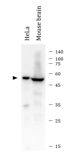
ARG57293 anti-Caspase 1 antibody WB image
Western blot: 10 µg of HeLa and 20 µg of Mouse brain lysates stained with ARG57293 anti-Caspase 1 antibody at 1:1000 dilution.
-

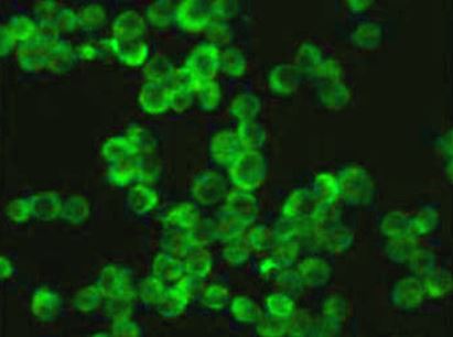
ARG57293 anti-Caspase 1 antibody ICC/IF image
Immunofluorescence: Raw264.7 cells stained with ARG57293 anti-Caspase 1 antibody at 1:100 dilution.
-

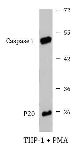
ARG57293 anti-Caspase 1 antibody WB image
Western blot: 25 µg of cell lysate from PMA treated THP-1 cells were stained with ARG57293 anti-Caspase 1 antibody at 1:1000 dilution.
-
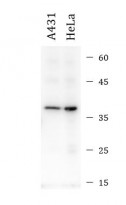
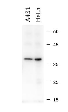
ARG66285 anti-IL1 beta antibody [SQab1748] WB image
Western blot: 20 μg of A431 and HeLa cell lysates stained with ARG66285 anti-IL1 beta antibody [SQab1748] at 1:4000 dilution.
-
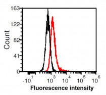
ARG66285 anti-IL1 beta antibody [SQab1748] FACS image
Flow Cytometry: HeLa cells stained with ARG66285 anti-IL1 beta antibody [SQab1748] at 1 µg/ml (red) and without antibody control (black).
-
ARG66285 anti-IL1 beta antibody [SQab1748] ICC/IF image
Immunofluorescence: MCF7 cells stained with ARG66285 anti-IL1 beta antibody [SQab1748] at 1:200 dilution.
-
ARG57293 anti-Caspase 1 antibody IHC-P image
Immunohistochemistry: Paraffin-embedded Mouse kidney tissue stained with ARG57293 anti-Caspase 1 antibody at 1:100 dilution.
-
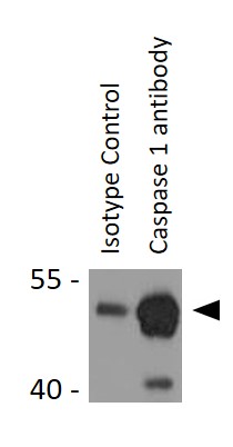
ARG57293 anti-Caspase 1 antibody IP image
Immunoprecipitation: 200 µg extracts of THP-1 cells Immunoprecipitated and stained with ARG57293 anti-Caspase 1 antibody at 1:1000 dilution.
-
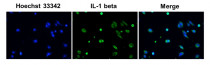
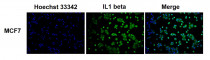
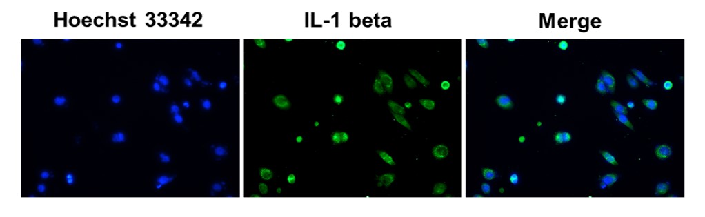
ARG66285 anti-IL1 beta antibody [SQab1748] ICC/IF image
Immunofluorescence: HeLa cells were fixed in 4% PFA, permeabilized with PBS containing 0.1% Triton X-100. Cells were stained with ARG66285 anti-IL1 beta antibody [SQab1748] (green) at 1:200 dilution, and cell nuclei were stained with Hoechst 33342 (blue).
-
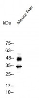
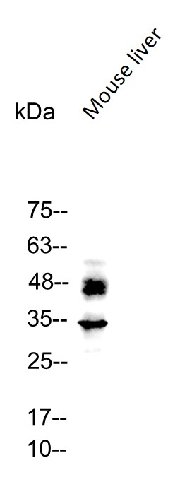
ARG66285 anti-IL1 beta antibody [SQab1748] WB image
Western blot: 50 µg of Mouse liver lysate stained with ARG66285 anti-IL1 beta antibody [SQab1748] at 1:5000 dilution.
-

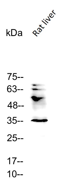
ARG66285 anti-IL1 beta antibody [SQab1748] WB image
Western blot: 50 µg of Rat liver lysate stained with ARG66285 anti-IL1 beta antibody [SQab1748] at 1:5000 dilution.


Specific References
Antioxidant and Anti-Inflammatory Effects of Turmeric Fermentation Liquid in Knee Osteoarthritis
ARG56625; IHC-P / Rat


Levistilide A ameliorates ischemic brain injury by suppressing the AIM2 inflammasome related pyroptosis
ARG41404: WB / Rat
NLRP3 inflammasome inhibits mitophagy during the progression of temporal lobe epilepsy
ARG41404; WB, IHC-P / Rat
Proanthocyanidins alleviate acute alcohol liver injury by inhibiting pyroptosis via inhibiting the ROS-MLKL-CTSB-NLRP3 pathway
ARG41404; WB / Mouse, Human ARG66285; WB / Mouse, Human
Protective effect of phosphoenolpyruvate carboxykinase 1 on inflammation and fibrotic progression of IgA nephropathy
ARG66285; IHC-P / Human
IGFBP5 promotes EndoMT and renal fibrosis through H3K18 lactylation in diabetic nephropathy
ARG57293; WB / Mouse
Preclinical evaluation of a novel antibody–drug conjugate OBI-992 for Cancer therapy
ARG62347: WB / Human
RNF167 mediates atypical ubiquitylation and degradation of RLRs via two distinct proteolytic pathways
ARG62347; WB / Human
Mechanisms of apigenin in mitigating APAP-induced hepatocyte pyroptosis and liver injury via TRPV4 activation and ESCRT-mediated membrane repair
ARG41404; WB / Mouse
Arctiin Mitigates Neuronal Injury by Modulating the P2X7R/NLPR3 Inflammasome Signaling Pathway
ARG57293; WB / Mouse
Inactivated SARS-CoV-2 induces acute skeletal muscle damage in human K18-hACE2 transgenic mice
ARG41404; WB / Mouse
Endophilin A2 controls touch and mechanical allodynia via kinesin-mediated Piezo2 trafficking
ARG62347: WB / Mouse
3-MCPD Induces Renal Cell Pyroptosis and Inflammation by Inhibiting ESCRT-III-Mediated Cell Repair and Mitophagy
ARG66285; WB / Rat
KDM3A Ablation Activates Endogenous Retrovirus Expression to Stimulate Antitumor Immunity in Gastric Cancer
ARG62347: WB / Human
Overexpression of EphB2 in the basolateral amygdala is crucial for inducing visceral pain sensitization in rats subjected to water avoidance stress
ARG62347: WB / Rat
黄芪甲苷预处理对大鼠肠缺血再灌注所致肺损伤的影响及其机制
ARG57293: WB / Rat
Discrepant Activation Pattern of Inflammation and Pyroptosis Induced in Dermal Fibroblasts in Response to Dengue Virus Serotypes 1 and 2 and Nonstructural Protein 1
ARG57293: WB / Human
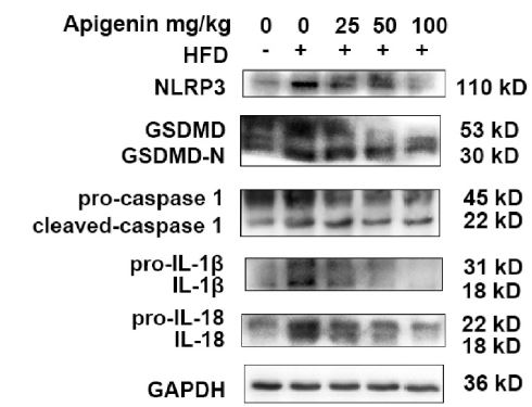
Apigenin Alleviated High-Fat-Diet-Induced Hepatic Pyroptosis by Mitophagy-ROS-CTSB-NLRP3 Pathway in Mice and AML12 Cells
ARG41404: WB / Mouse
Ginsenoside Rb1 alleviated concanavalin A-induced hepatocyte pyroptosis by activating mitophagy
ARG66285: WB / Human, Mouse
STING activation in cardiomyocytes drives hypertrophy-associated heart failure via NF-κB-mediated inflammatory response
ARG62347: WB / Human, Mouse
Ursolic acid induces apoptosis and pyroptosis in Reh cells by upregulating of the JNK signalling pathway based on network pharmacology and experimental validation
ARG57293: WB; ARG66285: WB / Human
Elaidic acid induced hepatocyte pyroptosis via autophagy-CTSB-NLRP3 pathway
ARG41404: WB / Mouse
Protective effect of ginsenoside Rh2 against Toxoplasma gondii infection-induced neuronal injury through binding TgCDPK1 and NLRP3 to inhibit microglial NLRP3 inflammasome signaling pathway
ARG57293: WB / Mouse
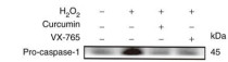
Curcumin improves the function of umbilical vein endothelial cells by inhibiting H2O2‑induced pyroptosis
ARG41404, ARG66285: WB; ARG57293:IF, WB / Human
Curcumin improves the function of umbilical vein endothelial cells by inhibiting H2O2 induced pyroptosis
ARG57293:ICC/IF / Human
Role of P2X7R in eosinophilic and non‑eosinophilic chronic rhinosinusitis with nasal polyps.
ARG66285: WB / Human
Curcumin Ameliorates White Matter Injury after Ischemic Stroke by Inhibiting Microglia/Macrophage Pyroptosis through NF- κ B Suppression and NLRP3 Inflammasome Inhibition
ARG41404: WB / Mouse
Aronia Melanocarpa Polysaccharide Ameliotates Inflammation and Aging in Mice by Modulating AMPK/SIRT1/NF-κB Signaling Pathway and Gut Microbiota
ARG41404: WB; ARG66285: WB; ARG57293: WB / Mouse
Abscisic acid ameliorates oxidative stress, inflammation, and apoptosis in thioacetamide-induced hepatic fibrosis by regulating the NF-кB signaling pathway in mice.
ARG66285: WB / Mouse


