ARG30323
Inflammation Antibody Panel
Component
| Cat No | Component Name | Host clonality | Reactivity | Application | Package |
|---|---|---|---|---|---|
| ARG56509 | anti-iNOS antibody | Rabbit pAb | Hu, Mamm, Ms, Rat | ICC/IF, IHC-P, IHC-Fr, IP, WB | 50 μl |
| ARG56491 | anti-COX2 antibody | Rabbit pAb | Gpig, Hu, Mk, Ms, Rb, Rat, Sheep | ICC/IF, IHC-P, WB | 50 μl |
| ARG51518 | anti-NFkB p65 phospho (Ser536) antibody | Rabbit pAb | Hu, Ms, Rat | ICC/IF, IHC-P, WB | 20 μl |
| ARG65683 | anti-beta Actin antibody | Rabbit pAb | Hu, Ms, Rb, Rat, Sheep | IHC-P, WB | 20 μg |
| ARG65351 | Goat anti-Rabbit IgG antibody (HRP) | Goat pAb | Rb | ELISA, IHC-P, WB | 50 μl |
Overview
| Product Description | Inflammation Antibody Panel is an all-in-one solution to make inflammation research easy and economic. It is ideal for studying inflammation in cultured cells. This antibody panel comprises the antibodies against key inflammatory mediators/markers iNOS and COX-2 and antibody against Ser536-phosphorylated NFkB p65 that is an NFkB activation marker in response to either LPS- or TNF alpha-induced inflammation. Moreover, the most suitable loading control beta-Actin antibody and the compatible secondary antibody are included in this panel. All the antibodies in this panel have excellent performance for not only WB but also more applications on multiple species. Related news: Inflammation antibody panels are released Exploring Antiviral Immune Response |
|---|---|
| Target Name | Inflammation |
| Alternate Names | Inflammation antibody; NFkB p65 phospho (Ser536) antibody; COX2 antibody; iNOS antibody; beta Actin antibody |
Properties
| Storage Instruction | For continuous use, store undiluted antibody at 2-8°C for up to a week. For long-term storage, aliquot and store at -20°C or below. Storage in frost free freezers is not recommended. Avoid repeated freeze/thaw cycles. Suggest spin the vial prior to opening. The antibody solution should be gently mixed before use. |
|---|---|
| Note | For laboratory research only, not for drug, diagnostic or other use. |
Bioinformation
| Gene Full Name | Antibody Panel for Inflammation |
|---|---|
| Highlight | Related products: anti-iNOS antibody; anti-COX2 antibody; Inflammation antibodies; Inflammation Duos / Panels; |
Images (27) Click the Picture to Zoom In
-
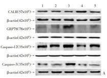
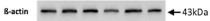
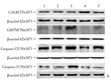
ARG65683 anti-beta Actin antibody WB image
Western blot: Human hepatic stellate cell stained with ARG20531 anti-BiP / GRP78 antibody, ARG54938 anti-Caspase 3 antibody, ARG55123 anti-Calreticulin antibody, ARG55177 anti-Caspase 12 antibody, and ARG65683 anti-beta Actin antibody. Secondary Antibody stained with ARG65351 Goat anti-Rabbit IgG antibody (HRP).
From DAI Linyu et al. Journal of Third Military Medical University (2021), doi: 10-16016-j-1000-5404-202012010, Fig. 4.
-

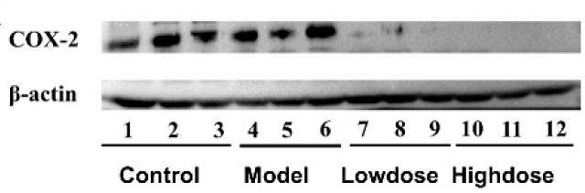
ARG56491 anti-COX2 antibody WB image
Western blot: Mouse stomach stained with ARG56491 anti-COX2 antibody.
From Zhu M et al. Foods (2025), doi: 10.3390/foods14091600, Fig. 4H.
-

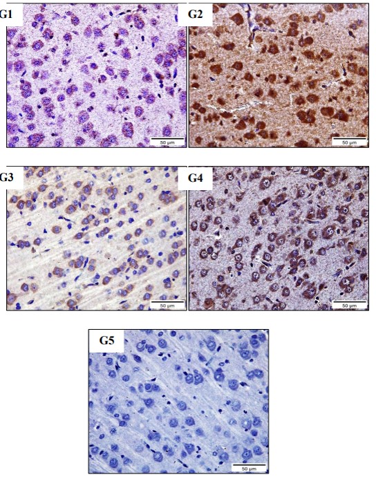
ARG56509 anti-iNOS antibody IHC-P image
Immunohistochemistry: Rat Brain stained with ARG56509 anti-iNOS antibody at 1:100 dilution.
From Abrar Roshdy Abouelkeir et al. European Chemical Bulletin,(2023) doi: 10.31838/ecb/2023.12.1.470, Fig. 6.
-

ARG51518 anti-NFkB p65 phospho (Ser536) antibody IHC-P image
Immunohistochemistry: Rat femoral head stained with ARG51518 anti-NFkB p65 phospho (Ser536) antibody at 1:300 dilution.
From Huihui Xu et al. Apoptosis. (2023), doi: 10.1007/s10495-023-01860-2, Fig. 6A.
-
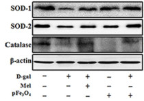
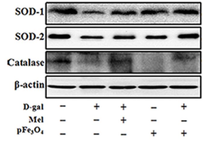
ARG65683 anti-beta Actin antibody WB image
Western blot: Mouse hippocampus stained with ARG22294 anti-SOD1 antibody , ARG54937 anti-SOD2 antibody and ARG65683 anti-beta Actin antibody.
From Zihao Xia et al. International Journal o f Molecular Sciences (2022), doi: 10.3390/ijms23126463, Fig. 6C.
-
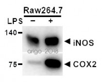
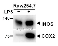
ARG56491 anti-COX2 antibody WB image
Western blot: 20 µg of Raw264.7 cells untreated or treated with LPS. The blots were stained with ARG55060 anti-iNOS antibody at 1:500 dilution and ARG56491 anti-COX2 antibody at 1:200 dilution.
-

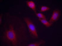
ARG56509 anti-iNOS antibody ICC/IF image (Customer's Feedback)
Immunofluorescence: RAW264.7 cells were fixed with 4% paraformaldehyde for 15 min at RT, permeabilized with 0.1% Triton X-100 then blocked with 2% albumin for 60 min at RT. Cells were stained with ARG56509 anti-iNOS antibody (green) at 4°C. DAPI (blue) was used as the nuclear counter stain.
-
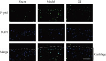
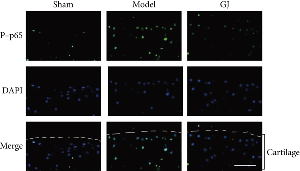
ARG51518 anti-NFkB p65 phospho (Ser536) antibody IHC-P image
Immunohistochemistry: Mouse tibial cartilage stained with ARG51518 anti-NFkB p65 phospho (Ser536) antibody.
From Congzi Wu et al. Biomed Res Int. (2022), doi: 10.1155/2022/9230784, Fig. 6. c.
-
ARG65683 anti-beta Actin antibody WB image
Western blot: C2C12 stained with ARG65683 anti-beta Actin antibody.
From Lin YH et al. Biomolecules (2021), doi: 10.3390/biom11111583, Fig. 1. C.
-

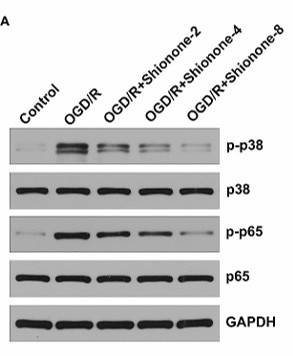
ARG51518 anti-NFkB p65 phospho (Ser536) antibody WB image
Western blot: SH-SY5Y stained with ARG51850 anti-p38 MAPK phospho (Thr180 / Tyr182) antibody, ARG55258 anti-p38 MAPK antibody, ARG51518 anti-NFkB p65 phospho (Ser536) antibody, and ARG57479 anti-NFkB p65 antibody.
From Zhou X et al. J Cardiothorac Surg (2024), doi: 10.1186/s13019-024-02938-x, Fig. 4. A.
-
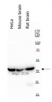
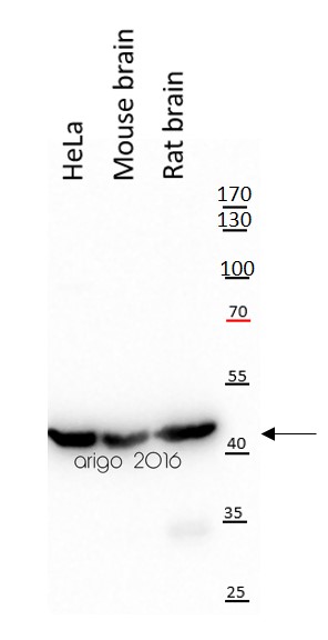
ARG65683 anti-beta Actin antibody WB image
Western blot: 20 µg of HeLa, Mouse brain and Rat brain lysates stained with ARG65683 anti-beta Actin antibody at 1:10000 dilution.
-
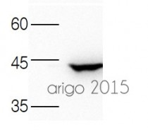
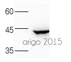
ARG65683 anti-beta Actin antibody WB image
Western blot: 30 µg of 293T lysate stained with ARG65683 anti-beta Actin antibody at 1:3000 dilution.
-
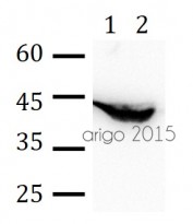
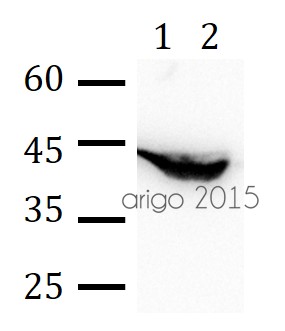
ARG65683 anti-beta Actin antibody WB image
Western blot: 30 µg of 1) Rat brain, and 2) Mouse liver lysate stained with ARG65683 anti-beta Actin antibody at 1:3000 dilution.
-
ARG56509 anti-iNOS antibody WB image
Western blot: Rat Aortic stained with ARG56509 anti-iNOS antibody at 1:1000 dilution.
From Wahid Shah et al. Sci Rep. (2023), doi: 10.1038/s41598-023-43786-4, Fig. 2. C.
-
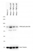
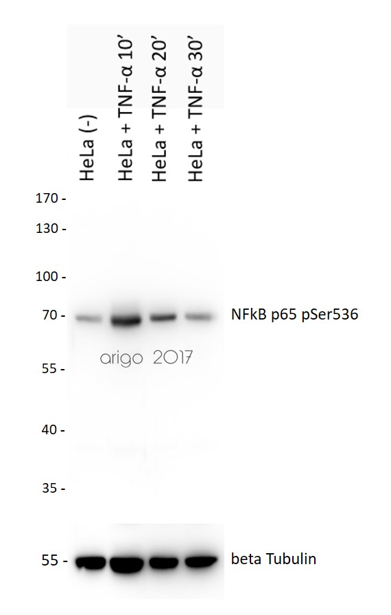
ARG51518 anti-NFkB p65 phospho (Ser536) antibody WB image
Western blot: 20 µg of HeLa cells untreated or treated with TNF-alpha at 10, 20 or 30 min. The blots were stained with ARG51518 anti-NFkB p65 phospho (Ser536) antibody at 1:500 dilution.
-
ARG51518 anti-NFkB p65 phospho (Ser536) antibody WB image
Western blot: Mouse heart stained with ARG51518 anti-NFkB p65 phospho (Ser536) antibody.
From Zhang J et al. Frontiers in Pharmacology (2022), doi: 10.3389/fphar.2022.890202, Fig. 4. E.
-
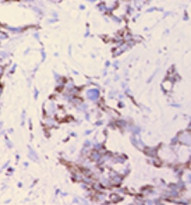
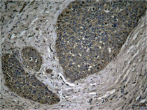
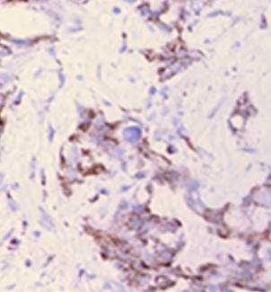
ARG56509 anti-iNOS antibody IHC-P image
Immunohistochemistry: Paraffin-embedded Human pancreatic ductal adenocarcinoma stained with ARG56509 anti-iNOS antibody.
-
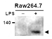
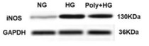
ARG56509 anti-iNOS antibody WB image
Western blot: Raw264.7 cells untreated or treated with LPS. 20 µg of cell lysates stained with ARG56509 anti-iNOS antibody at 1:400 dilution.
-

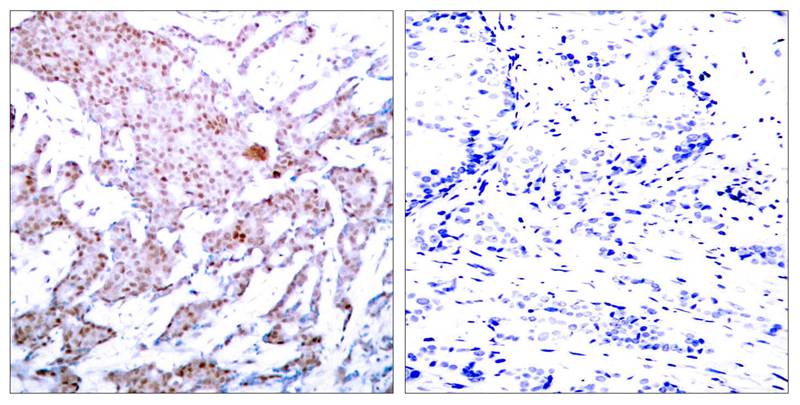
ARG51518 anti-NFkB p65 phospho (Ser536) antibody IHC-P image
Immunohistochemistry: Paraffin-embedded Human breast carcinoma tissue stained with ARG51518 anti-NFkB p65 phospho (Ser536) antibody (left) or the same antibody preincubated with blocking peptide (right).
-
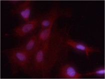
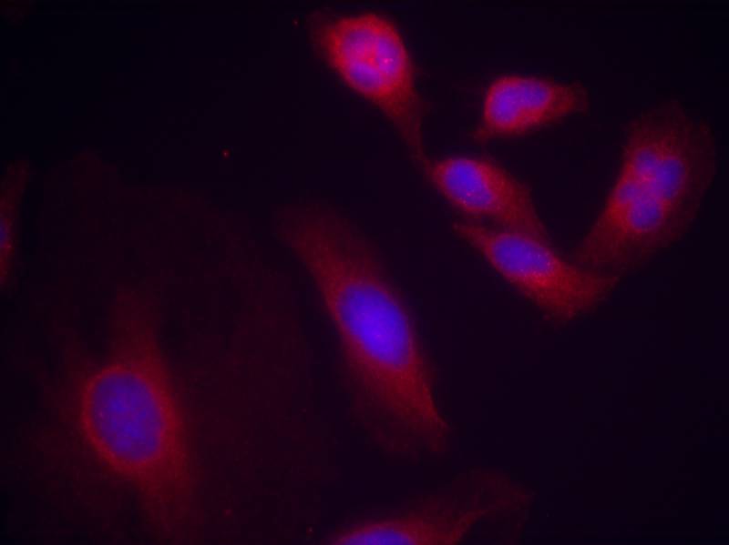
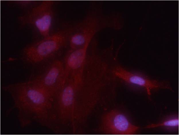
ARG51518 anti-NFkB p65 phospho (Ser536) antibody ICC/IF image
Immunofluorescence: methanol-fixed HeLa cells stained with ARG51518 anti-NFkB p65 phospho (Ser536) antibody.
-
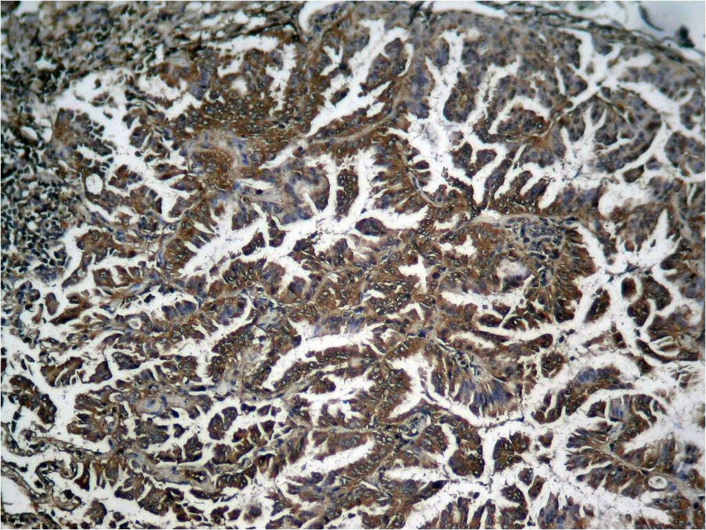
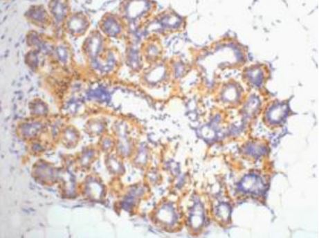
ARG51518 anti-NFkB p65 phospho (Ser536) antibody IHC-P image
Immunohistochemistry: Paraffin-embedded Human breast carcinoma tissue stained with ARG51518 anti-NFkB p65 phospho (Ser536) antibody.
-

ARG51518 anti-NFkB p65 phospho (Ser536) antibody IHC-P image
Immunohistochemistry: Paraffin-embedded Human Lung carcinoma tissue stained with ARG51518 anti-NFkB p65 phospho (Ser536) antibody.
-
ARG51518 anti-NFkB p65 phospho (Ser536) antibody ICC/IF image
Immunofluorescence: methanol-fixed MEF cells stained with ARG51518 anti-NFkB p65 phospho (Ser536) antibody.
-

ARG65683 anti-beta Actin antibody IHC-P image
Immunohistochemistry: Human ovary tissue stained with ARG65683 anti-beta Actin antibody at 1:200 dilution.
-

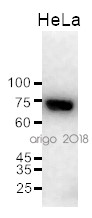
ARG56491 anti-COX2 antibody WB image
Western blot: 20 µg of HeLa cell lysate stained with ARG56491 anti-COX2 antibody at 1:200 dilution.
-
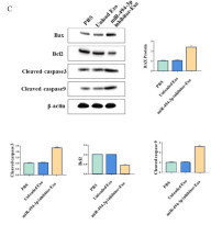
ARG65351 Goat anti-Rabbit IgG antibody (HRP) WB image
Western blot: Gastric cancer cells stained with ARG66247 anti-Bax antibody, ARG55188 anti-Bcl 2 antibody, ARG57512 anti-Caspase 3 (cleaved) antibody and ARG62346 anti-beta Actin antibody [BA3R].
Secondary Antibody stained with ARG65351 Goat anti-Rabbit IgG antibody (HRP).From Limin Zhang et al. Heliyon (2024), doi: 10.1016/j.heliyon.2024.e30803, Fig. 4. C.
-

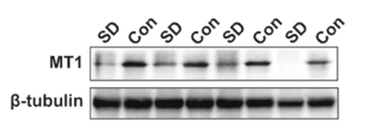
ARG65351 Goat anti-Rabbit IgG antibody (HRP) WB image
Western blot: Rat placental stained with ARG57589 anti-MTNR1A antibody at 1:1000 dilution, ARG65351 Goat anti-Rabbit IgG antibody (HRP) at 1:5000 dilution.
From Jinzhi Li et al. J Reprod Immunol. (2023), doi: 10.1016/j.jri.2023.104166, Fig. 2.B.


Specific References
Bioactive Compounds of Momordica charantia L. Downregulate the Protein Expression of ACE2 and TMPRSS2 In Vivo and In Vitro
ARG65351; WB /


Methylophiopogonanone a attenuates pulmonary fibrosis by inhibiting SPP1-mediated macrophage polarization via the PI3K/Akt pathway
ARG56509; IHC-P / Mouse
Mechanical and interfacial properties of zwitterionic hydrogels via a high-entanglement network design: a study of monomer and crosslinker synergy
ARG65351: FuncSt /
Investigation of the regulatory effect of overexpressed Ptpn2 on SiO2-mediated mouse alveolar macrophages based on iTRAQ technology
ARG51518: WB / Mouse
Soybean Trypsin Inhibitor Possesses Potency Against SARS-CoV-2 Infection by Blocking the Host Cell Surface Receptors ACE2, TMPRSS2, and CD147
ARG65351; WB /
Reduced endometrial glycolysis concomitant with increased lesional fibrosis in patients with adenomyosis who complained of heavy menstrual bleeding
ARG65351; WB /
Progressively diminished estrogen signaling concordant with increased fibrosis in ectopic endometrium
ARG65351; WB /
Necroptosis of hippocampal neurons in paclitaxel chemotherapy-induced cognitive impairment mediates microglial activation via TLR4/MyD88 signaling pathway
ARG56509; IHC-Fr / Mouse
Selenium Mitigates Caerulein and LPS-induced Severe Acute Pancreatitis by Inhibiting MAPK, NF-κB, and STAT3 Signaling via the Nrf2/HO-1 Pathway
ARG51518; WB / Mouse
Structure-based design of potent and selective inhibitors targeting RIPK3 for eliminating on-target toxicity in vitro
ARG65683; WB / Human, Mouse
Effect of Elaeagnus angustifolia Honey in the Protection Against Ethanol-Induced Chronic Gastric Injury via Counteracting Oxidative Stress, Interfering with Inflammation and Regulating Gut Microbiota in Mice
ARG56491; WB / Mouse
Pdlsc-Cm Targets Hapln1 To Reduce Root Resorption After Replantation
ARG56509; IHC-P, WB / Rat
Melatonin receptor 1A/B knockout accelerates atrial aging in mice
ARG56509; IHC-P / Mouse
Identification of RIPK3 as a target of flavonoids for anti-necroptosis in vitro
ARG65683; WB / Mouse
Effect of macrophage-to-myofibroblast transition on silicosis
ARG56509: IHC-P,WB / Rat
Protein degradation of antizyme depends on the N-terminal degrons
ARG65683; WB / Human
Chronic intermittent hypobaric hypoxia alleviates early-stage posttraumatic osteoarthritis via NF-κB/Nrf2 pathway in mice
ARG56509; WB / Mouse
Overexpression of ZNF468 promotes esophageal squamous cell carcinoma progression via the AKT/mTOR pathway
ARG65351; WB /
Aquaporin-9 Aggravates Lipopolysaccharides Induced Acute Lung Injury Via Facilitating M1-Like Macrophage Polarization
ARG56509: IHC-P / Mouse
Chronic intranasal oxytocin alleviates cognitive impairment and reverses oxytocin signaling upregulation in MK801-induced mice
ARG65351; WB /
Melatonin alleviates aging-related heart failure through melatonin receptor 1A/B knockout in mice
ARG56509: IHC-P / Mouse
Discovery and mechanism of anti-hypertensive effect of a novel tripeptide (SYP) from medicinal fungus Ganoderma lingzhi
ARG65683: WB / Human
Engineered extracellular vesicles with polypeptide for targeted delivery of doxorubicin against EGFR‑positive tumors
ARG65683: WB / Human
Anti-CTLA-4 antibody self-presented dendritic cell nanovesicles boost the immunotherapy of hepatocellular carcinoma after microwave ablation
ARG65351; WB /
Facile and rapid fabrication of a novel 3D-printable, visible light-crosslinkable and bioactive polythiourethane for large-to-massive rotator cuff tendon repair
ARG56509: IHC-P / Mouse
Intravital microscopic thermometry of rat mammary epithelium by fluorescent nanodiamond
ARG51518: IHC-P / Rat
Echinacoside exerts neuroprotection via suppressing microglial α-synuclein/TLR2/NF-κB/NLRP3 axis in parkinsonian models
ARG51518: WB / Mouse
Shionone relieves oxygen-glucose deprivation/reoxygenation induced SH-SY5Y cells injury by inhibiting the p38 MAPK/NF-κB pathway
ARG51518: WB / Human
The role of phlorizin liposome-embedded oxidized sodium alginate/carboxymethyl chitosan in diabetic wound healing
ARG51518; WB / Mouse
Banxia-Houpu decoction inhibits iron overload and chronic intermittent hypoxia-induced neuroinflammation in mice
ARG56509: WB, IHC-P / Mouse
Identification and experimental verification of immune-related hub genes in intervertebral disc degeneration
ARG56509: ICC/IF / Human
Calycosin Induces Ferroptosis by SLC7A11 Through the PI3K/Akt Pathway in Acute Myelocytic Leukemia
ARG65351; WB /
Targeting VCP potentiates immune checkpoint therapy for colorectal cancer
ARG65683: WB / Mouse
TNFAIP3 interacting protein 2 relieves lipopolysaccharide (LPS)-induced inflammatory injury in endometritis by inhibiting NF-kappaB activation
ARG51518: WB / Human
Polydatin improves vascular endothelial function by maintaining mitochondrial homeostasis under high glucose conditions
ARG56509: WB / Rat
Anti-Inflammatory Actions of G-Protein-Coupled Estrogen Receptor 1 (GPER) and Brain-Derived Estrogen Following Cerebral Ischemia in Ovariectomized Rats
ARG56509: WB, IHC-Fr / Rat
Aucubin Alleviates Intervertebral Disc Degeneration by Repressing NF-κB-NLRP3 Inflammasome Activation in Endplate Chondrocytes
ARG51518: IHC-P / Mouse
Elucidation of the Underlying Mechanism of Gujian Oral Liquid Acting on Osteoarthritis through Network Pharmacology, Molecular Docking, and Experiment
ARG51518: IHC-P / Mouse
N-Butanol Extract of Modified You-Gui-Yin Attenuates Osteoclastogenesis and Ameliorates Osteoporosis by Inhibiting RANKL-Mediated NF-κB Signaling
ARG51518: IHC-P / Mouse
Paclitaxel induces cognitive impairment via necroptosis, decreased synaptic plasticity and M1 polarisation of microglia
ARG56509: IHC-Fr / Mouse
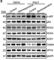
Neohesperidin Protects Angiotensin II-Induced Hypertension and Vascular Remodeling.
ARG51518: WB / Mouse
Dual keratinocyte-attachment and anti-inflammatory coatings for soft tissue sealing around transmucosal oral implants
ARG56509: ICC/IF / Mouse
GNL3 is an evolutionarily conserved stem cell gene influencing cell proliferation, animal growth and regeneration in the hydrozoan Hydractinia
ARG51518: WB / Human
Berberine ameliorates mesenteric vascular dysfunction by modulating perivascular adipose tissue in diet-induced obese in rats
ARG56509: IHC-P / Rat
Glycolytic Reprogramming in Silica-Induced Lung Macrophages and Silicosis Reversed by Ac-SDKP Treatment
ARG56509: WB / Rat
Chlorogenic Acid retards cartilaginous endplate degeneration and ameliorates intervertebral disc degeneration via suppressing NF-κB signaling.
ARG51518: IHC-P / Mouse
Hepatoma-derived growth factor participates in concanavalin A-induced hepatitis.
ARG56491: WB, IHC-P / Mouse, Rat
Benzophenone and Benzoylphloroglucinol Derivatives from Hypericum sampsonii with Anti-Inflammatory Mechanism of Otogirinin A.
ARG56491: WB / Mouse
Baihe Wuyao decoction ameliorates CCl 4-induced chronic liver injury and liver fibrosis in mice through blocking TGF-β1/Smad2/3 signaling, anti-inflammation and anti-oxidation effects.
ARG56509: WB / Mouse
A free-standing multilayer film as a novel delivery carrier of platelet lysates for potential wound-dressing applications.
ARG56509: IHC-P / Rat
HYCO-3, a dual CO-releaser/Nrf2 activator, reduces tissue inflammation in mice challenged with lipopolysaccharide.
ARG56509: IHC-P / Mouse
Distribution and proportion of M1/M2 macrophages in periodontal tissues in rats with and without periodontitis
ARG56509: IHC-P / Rat


