LAG-3, TIGIT, and TIM-3: key drivers of T cell exhaustion
LAG-3, TIGIT, and TIM-3: key drivers of T cell exhaustion
|
|
|
|
|
|
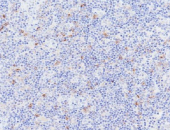
![Immunohistochemistry (IHC-P) staining of human lymphoma tissue using anti-CD223 / LAG-3 antibody [SQab22258], FFPE section with Tris-EDTA antigen retrieval (pH 9.0).](/files/editor/images/ARG66930%20anti-CD223%20_%20LAG3%20antibody%20SQab22258%20IHC-P.jpg)
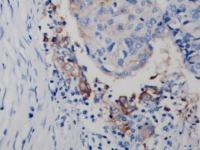
![Immunohistochemistry: Human tonsil stained with anti-TIGIT antibody [SQab30360]](/files/editor/images/ARG67087%20anti-TIGIT%20antibody%20%5BSQab30360%5D%20IHC-P.jpg)
|
|
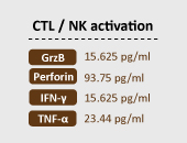
More research tools for immunosuppression
|
COX2-PGE2 Axis |
IDO Pathway Tryptophan ELISA Kit (ARG80483) Kynurenine ELISA Kit (ARG82779)
|
Adenosine axis Adenosine Assay Kit (ARG83372) |
|
|
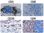
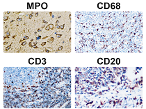
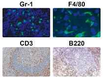
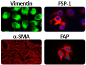
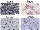
Solutions for TME visualization
|
|
You may be interested in...
- The best solution for PD-1/PD-L1 research
- Detecting exosomal PD-L1 secreted by cancer cells
- PD-1 ELISA Kits, excellent for preclinical studies or pharmatheutical development
